|
|
Esophageal Duplication Cyst
General Considerations
- Rare mediastinal mass
- Most are diagnosed in childhood
- May be simple epithelial cysts or duplications that contain muscle and submucosa but not epithelium
- Most (60%) occur in lower 1/3 of esophagus
- Most commonly associated with difficulty swallowing
- Occur because of maldevelopment of the posterior division of embryonic foregut
Clinical Findings
- May be asymptomatic and never diagnosed
- Symptoms are produced by compression of surrounding structures; larger cysts produce more symptoms
- Those that occur in cervical esophagus (20%) may produce respiratory problems most commonly
- Those that occur in mid-esophagus (20%) may cause retrosternal pain or dysphagia
- May present with hemorrhage if there is gastric mucosa in cyst
Imaging Findings
- Most develop on the right side of esophagus, posteriorly and inferiorly
- Conventional radiography
- Sharply-marginated, middle mediastinal soft tissue mass
- CT scans
- Well-marginated, usually round, oval or tubular-shaped, fluid-filled cystic structure
- Well-defined, thin wall
- Homogeneous water attenuation (0-20HU)
- No enhancement of cyst contents
- No infiltration of surrounding tissues
- Originates from esophagus
- MRI
- Typically dark on T1 and bright on T2-weighted images
Differential Diagnosis
- Bronchogenic cysts (may have identical appearance to esophageal cysts)
- Neurenteric cysts
- Pericardial cysts
- Cystic teratoma
Treatment
- Nearly ¾ with cysts will become symptomatic, so they are usually resected
- Simple cysts are enucleated
- Duplication cysts are excised
Complications
- Complication rates from surgery are very low
- Malignant degeneration is rare but reported
Prognosis
 
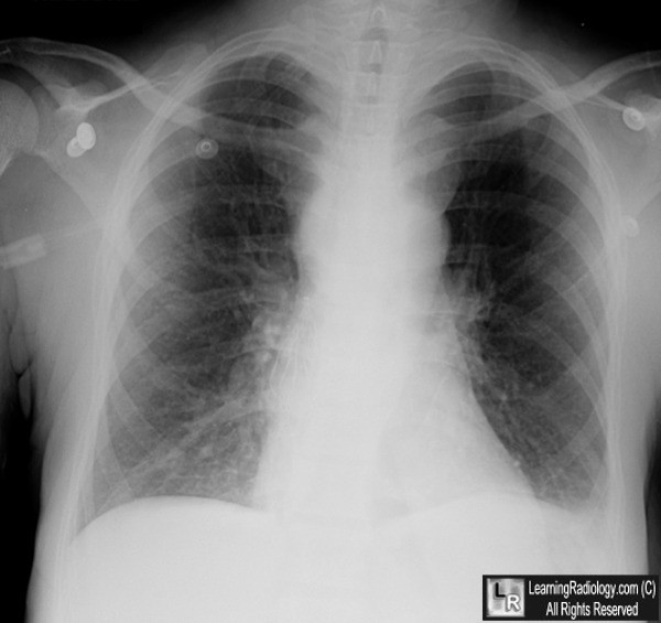
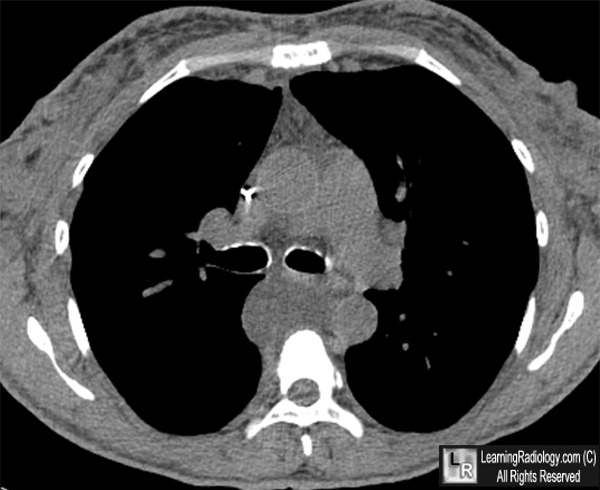
Esophageal Duplication Cyst. White arrows point to a middle mediastinal soft tissue mass with sharp
margins on the conventional frontal radiograph. On the CT scan, the white arrow points to a fluid-filled,
cystic mass to the right of the location of the esophagus. The mass measured water density.
For more information, click on the link if you see this icon 
For these same photos without the annotations, click here and here
Esophageal Cysts. eMedicine. Dale K Mueller, MD.
Imaging of Cystic Masses of the Mediastinum. Mi-Young Jeung, MD, Bernard Gasser, MD, Afshin Gangi, MD, PhD, Adriana Bogorin, MD, Dominique Charneau, MD, Jean Marie Wihlm, MD, Jean-Louis Dietemann, MD and Catherine Roy, MD. RadioGraphics, 22, S79-S93.
|
|
|
