|
|
Caseous Calcification of the Mitral Annulus
CCMA
General Considerations
- Mitral annular calcification is found in 10% of patients > than 50 years of age, most often in women
- It is a chronic degenerative process almost always asymptomatic
- Represents deposition of calcium between the basal infero-lateral ventricular wall and posterior leaflet of mitral valve
- Caseous calcification of the mitral annulus is an uncommon variant of mitral annular calcification with a characteristic appearance
- Represents a mixture of calcium, cholesterol and fatty acids
Clinical Findings
- Patients are usually asymptomatic
- Most are older and almost all have systemic hypertension
Imaging Findings
- Large, spherical calcification that may have sonolucent center on echo
- On MRI, it is of low signal intensity on both T1 and T2-weighted images before and after contrast.
- Usually located on the posterior mitral leaflet
- Contains putty-like, “toothpaste-like” material surrounded by a calcified shell
Differential Diagnosis
- Benign prognosis not to be mistaken for tumor which is in the differential diagnosis
Prognosis
- There are reported cases in which the calcification spontaneously disappeared on follow-up and in another case presumably resolved related to hemodialysis
 
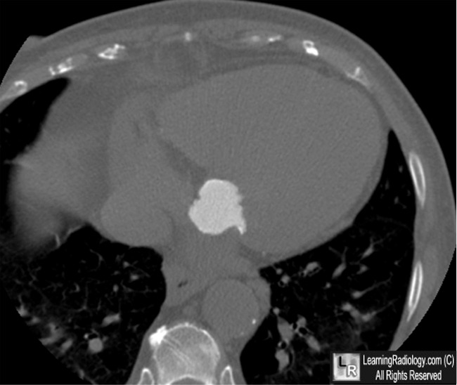
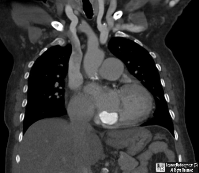
Caseous Calcification of the Mitral Annulus. Post-contrast enhanced CT axial and coronal scans
through the heart show a large, lobulated calcification at the site of the mitral valve (white arrows)
representing the characteristic appearance of caseous calcification of the mitral annulus.
For more information, click on the link if you see this icon 
For these same photos without the annotations, click here and here
Caseous Calcification of the Mitral Valve Ring. Marcu , C, Ghantous, A and Prokop, E. Lung and Circulation. Volume 15, Issue 3, June 2006, Pages 187-188.
Caseous Calcification of the Mitral Annulus. H Arora; P Madan; L Simpson and R Stainback. Tex Heart Inst J. 2008; 35(2): 211–213. 2008
Cardiovascular magnetic resonance features of caseous calcification of the mitral annulus. L Monti; E Renifilo; M Profili and L Balzarini. Cardiovasc Magn Reson. 2008; 10(1): 25.
|
|
|
