|
|
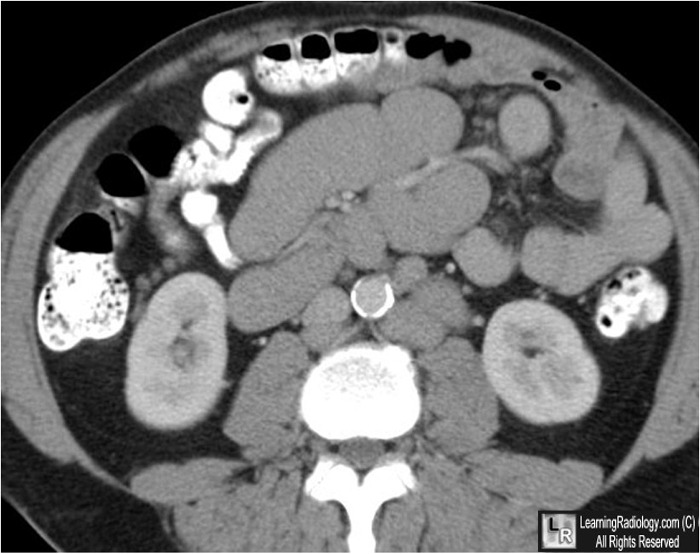
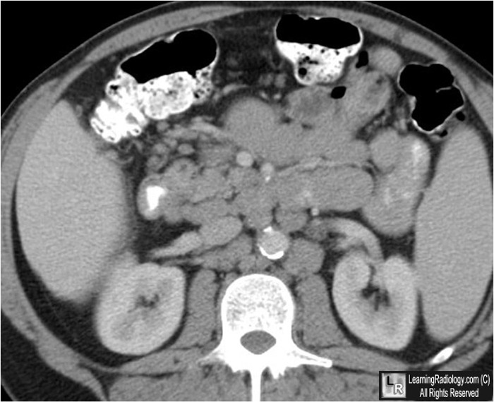
Sandwich Sign
Hamburger Sign
General Considerations
- CT appearance of mesenteric fat and vessels "sandwiched" between two nodular layers of mesenteric lymphadenopathy
- Highly suggestive of mesenteric lymphoma
- Usually non-Hodgkin's variety (NHL)
- Mesenteric masses occur in 30-50% of patients with NHL
- Frequently associated with retroperitoneal adenopathy as well
- Sandwich sign may also be seen in patients with post-transplantation lymphoproliferative disorder
 
Sandwich Sign. Axial contrast-enhanced CT images of the abdomen and pelvis
demonstrate lobulated, lymphadenopathy (white arrows) "sandwiching" mesenteric vessels and fat (red arrows) in a pattern suggestive of mesenteric lymphoma. There is also retroperitoneal adenopathy (black arrows).
For more information, click on the link if you see this icon 
For this same photo without the annotations, click here and here
The Sandwich Sign. Hardy, S. Radiology, 226, 651-652.
|
|
|
