|
|
Ventriculo-Peritoneal Shunt Complications
General Considerations
- Ventriculo-peritoneal (VP) shunts are used to relieve hydrocephalus
- Complication rates have been reported as high as 81% after 12 years
- Complications include
- Infections
- Shunt occlusion
- Overdrainage
Infections
- Most occur within 2 months of surgery
- Most infections are from direct spread
- Bacteria involved are
- Staphylococci (60%)
- Remaining 40% include coryneforms (Propionibacterium), streptococci, enterococci, aerobic Gram-negative rods, and yeasts.
- In late infections, suspect Gram-negative bacilli infection from another site, such as abdominal infection
- Risk factors for shunt infections include:
- Young age
- Poor condition of the skin
- Intercurrent systemic infections
- Previous CNS infections
- Multiple revisions
- Types of infection
- Wound infection
- Meningitis/ventriculitis
- Peritonitis
- Infected shunt apparatus
Mechanical complications
- Occlusion
- About 50% of shunt complications in pediatric series
- Occlusion can occur at three different levels
- Proximal catheter
- Valve system
- Distal catheter.
- Risk for occlusion is higher during immediate postoperative period and decreases progressively with time
- Diagnosis is confirmed with increased ventricular size on CT scan
- Disconnection and fracture
- Shunt disconnection/fracture comprises 2nd most common cause of mechanical shunt malfunction
- Disconnection is defined as loss of continuity of shunt at normal connecting points between catheters, valves, and/or connectors
- Fracture is actual breakage of the catheter with separation of segments
- Associated factors for broken shunts are
- Growth spurts
- Aging, brittle or partially calcified shunt
- Multiple proximal revisions
- Local trauma to shunt
- Athletic activity without history of direct trauma
- Post scoliosis correction
- Shunt design (multiple shunt pieces have more risk of disconnecting
- Most common location of breakage is in neck, followed by the scalp either proximal or distal to valve or connecting devices
- Patients with broken shunts may present with
- Signs of increased intracranial pressure
- Pain
- Fluid collections along shunt tract and/or a palpable gap
- Asymptomatic patients are usually diagnosed as an incidental radiological finding or during follow-up visits
- Diagnosis of disconnection or fracture is confirmed on conventional radiographs (shunt series which show entire length of shunt from skull to abdomen).
- CT scan can show increase in ventricular size or no change
- Migration
- Shunt must be pulled and have ability to move in the subcutaneous tissue
- Loose or improper connection may allow catheters to migrate
- Overdissection of the subcutaneous tissue also predisposes to migration
- Valves not fixed in the subcutaneous tissue pose a high risk to migrate into the distal site or, retrograde into ventricles. Cylindrical valves are more prone to migration
- Migration may occur early after shunting or over time
- Patients present similar to disconnection/fracture
Improper placement
- Shunt can be improperly placed at level of ventricles or drainage cavity
- Signs and symptoms of improper placement similar to shunt occlusion
- Appear in early postoperative period
- Once suspected, shunt series and head CT scan are used to confirm diagnosis
- Lateral abdominal film may show an extra-peritoneal distal catheter
Overdrainage
- Overdrainage can occur normally with postural changes, REM sleep, straining
- Overdrainage can cause
- Symptomatic orthostatic hypotension
- Subdural CSF collections
- Slit ventricle syndrome
- Craniosynostosis
- Loculation of the ventricles.
- Risk of overdrainage can be minimized by increasing opening pressure of valve (programmable valves)
- Or by adding a siphon-resistive device to the system
- Or by using a flow-regulating device
Complications related to specific types of shunts
Ventriculopleural shunts
- Pleural effusions are most common complication
- Usually small and do not require treatment
- Pleural empyemas are uncommon
Ventriculoatrial shunts
- Improper placement is one of most common complications of VA shunt
- Bacterial endocarditis is most important infectious complication
- Nephritis has also been reported as a complication of VA shunts
Lumboperitoneal shunts
- Acquired Chiari malformation with tonsillar herniation
- Other complications include scoliosis and hyperlordosis, limited spinal flexion, transient back pain, sciatica, transient neck pain, lower limb neurological changes, arachnoiditis, and rarely, migration of the LP shunt

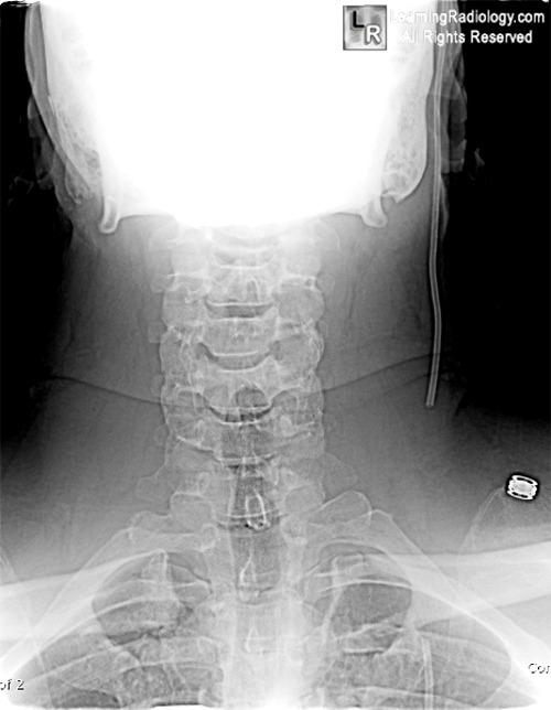
Fractured VP Shunt. Frontal radiograph of the cervical spine (edge-enhanced) shows a fracture of the ventriculo-peritoneal shunt drainage tube (black and white arrows) with caudal retraction of the distal fragment.
For more information, click on the link if you see this icon 
For this same photo without the annotations, click here
Ventricular Shunts. Ahmed, K., Ghotme, G. and Drake, J. Hospital for Sick Children, University of Toronto Toronto, Canada
|
|
|
