Wegener's Granulomatosis
Submitted by Denise Drohobyczer, MSIV |
General Considerations
- Hallmarks
- Small to medium sized systemic vasculitis
- Granulomatous inflammation
- Necrosis
- High C-ANCA association (88%)
- Not a definitive marker for diagnosis
- Unknown etiology
- Incidence of 1 case per 30,000
- Affects predominantly whites (91%)
- M>F
- Mean age at diagnosis = 45
Clinical Findings
- Classic Form
- Granulomatous inflammation of upper and lower respiratory tracts and kidneys.
- Limited Form
- Isolated to upper respiratory tract or lungs.
Sites of Involvement:
- Lung (>90%)
- Renal (75%)
- Rapidly progressing glomerulonephritis, chronic renal failure
- Trachea (15-60%)
- Other
- Paranasal sinuses, nose (saddle nose deformity)
- Ears, eyes, oral cavity
- Skin, joints, nervous system
- Rarely
- Heart, GI tract and brain
- Can potentially involve any organ in the body
Presentation:
- Nonspecific signs and symptoms
- Systemic complaints
- Fever, malaise, arthralgias and weight loss
- Upper and lower respiratory tract symptoms (85%)
- Upper respiratory
- Rhinitis is often the first symptom
- Mucosal ulcerations, epistaxis, refractory otitis media and sinusitis, hearing loss
- Lower respiratory
- Cough, dyspnea and hemoptysis
- Tracheal involvement
- Nonspecific laboratory findings
- Leukocytosis, thrombocytosis, elevated ESR and CRP, normochromic normochromic anemia
- Proteinuria, elevated BUN and creatinine, urine sediment with red cell casts
Diagnosis:
- American College of Rheumatology Classification
- (2 out of 4 criteria is 88% sensitive and 92% specific for the diagnosis)
- Nasal or oral inflammation
- Abnormal chest X-ray
- Urinary sediment
- Biopsy
Imaging Findings
Conventional Radiography
- Pulmonary nodules
- Most common finding (40-70%)
- Typically multiple and bilateral with a tendency to cavitate (50%)
- Cavitary lesions may lead to atelectasis or pneumothorax
- Thick or thin walled, well or ill circumscribed
- Tendency to wax and wane
- Size varies (1.5-10cm)
- Air space consolidation
- Waxing and waning infiltrates
- May be mistaken for pneumonia
- Pulmonary hemorrhage or edema
- Hilar lymphadenopathy
- Normal in 20%
CT
- Useful in further defining extent of disease seen in plain film, and revealing lesions not seen on plain film including:
- Interstitial abnormalities
- Tracheobronchial Abnormalities
- Findings suggestive of vasculitis
Differential Diagnosis
For cavitary lung lesions
- Infarction
- Septic pulmonary emboli
- Carcinoma
- Infection
- Rheumatoid nodules
Prognosis and Treatment
- Mainstay
- Corticosteroids and cyclophosphamide
- Maintenance
- Methotrexate or Azathioprine
- Tracheostomy may be required for tracheal strictures
- Independent Risk factors of mortality
- Older age
- Absence of ear, nose and throat involvement
- Renal or Cardiac involvement
|
|
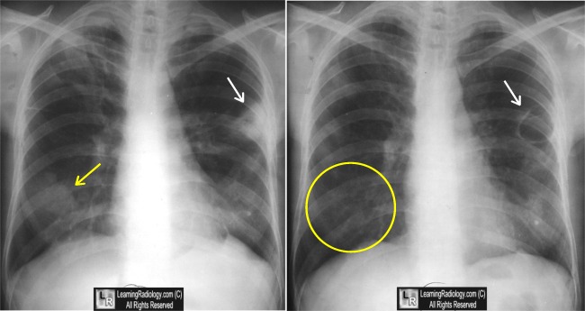
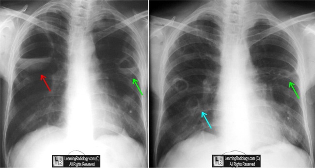
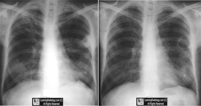
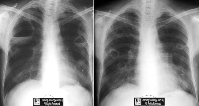
Wegener's Granulomatosis. Four images span approximately two years and show typical waxing and waning of pulmonary masses (white and green arrows), some of which cavitate (blue arrow), some of which disappear over the course of time (yellow circle).
For more information, click on the link if you see this icon 
For this same photo without the annotations, click here and here
|
| |
|
|
|
Ravenel, JG, MD; Irshad, A, MD. Wegener Granulomatosis, Thoracic. eMedicine
King, TE, MD. Respiratory tract involvement in Wegener's granulomatosis. UpToDate |
|