|
|
Right Middle Lobe Syndrome
General Considerations
- Definition varies
- Usually refers to any cause of recurrent or persistent atelectasis of the right middle lobe
- Occurs at all ages but most common at two peaks: in children and over 50
- Reportedly more common in women than men
- Originally thought to be due only to bronchial obstruction, but it is more often present with non-obstructive lesions
- Some believe the right middle lobe is more often involved because of its isolation and poor collateral flow from the upper and lower lobes
- Etiologies include
- Inflammatory disease is the most common etiology
- In children, it is most common in association with asthma
- Malignant tumors (22%0
- Bronchiectasis (15%)
- Tuberculosis (9%)
- Benign tumors (2%)
- In most cases, the etiology is never found
Clinical Findings
- Most often asymptomatic
- Cough
- Wheezing
- Dyspnea
Imaging Findings
- Right middle lobe atelectasis is usually easier to recognize on the lateral view than the frontal view, where it may produce very subtle findings
- Silhouetting of the right heart border on the frontal view by the adjacent un-aerated medial segment of the middle lobe
- If the atelectatic middle lobe swings upward and anteriorly, it may produce a wedge-shaped density on the frontal view with its base at the heart
- Depression of the minor fissure and elevation of the major fissure, especially well seen on the lateral view
- On the lateral view, the atelectatic lobe forms a triangular density with its apex at the hilum and its base more peripheral in the lung
- If there is a nodular density seen at the apex of the triangle on the lateral view, suspect a mass in the hilum producing the atelectasis
- Elevation of the right hemidiaphragm may occur
- CT is useful in excluding a cause of bronchial obstruction
- Middle lobe bronchus enters consolidated lobe in the posteromedial corner
Differential Diagnosis
Treatment
- Depends on etiology
- Most are treated with medical therapy alone
Prognosis
- Cases in about 1/3 of pediatric patients resolve after bronchoscopy


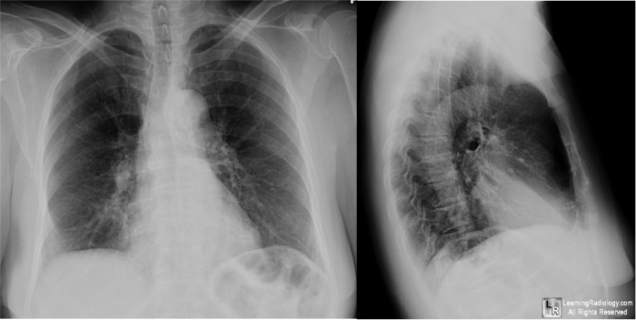
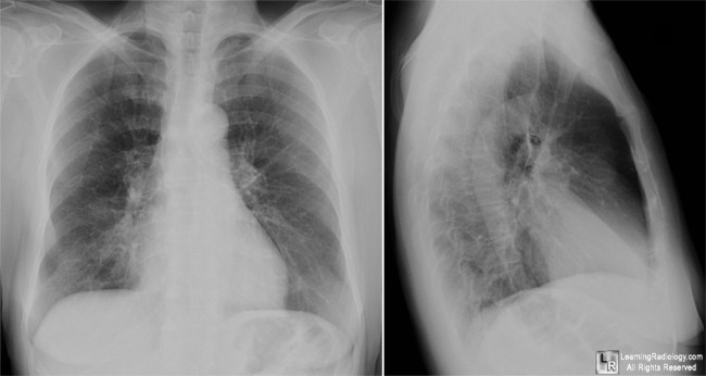
Right Middle Lobe Syndrome. Chest images at top are taken 3 months before images on bottom.
Both show middle lobe atelectasis with silhouetting of the right heart border on the frontal view (white arrows)
and a wedge-shaped density on the lateral with a depressed minor fissure (yellow arrows).
For more information, click on the link if you see this icon 
For this same photo without the annotations, click here and here
Middle Lobe Syndrome Due To Tuberculous Etiology: A Series Of 12 Cases.
Gupta1, P; Gupta, K and Agarwal, D. Indian J Tuberc 2006; 53:104-108
Diagnostic thoracic imaging By Wallace T. Miller. McGraw-Hill Professional, 2006
|
|
|
