|
|
Pelvic Lipomatosis
General Considerations
- Rare disease characterized by an excess deposition of fat in the pelvis surrounding the bladder and rectum
- Most common in African-Americans (66%) with an overwhelming male predominance (94%)
- Cause is not known but, it has been linked to obesity
- Most patients are 20-50 years of age
Clinical Findings
- From compression on the bladder (more common) and rectum
- Nocturia
- Dysuria
- Constipation
- Ribbon-like stools
- Lower extremity swelling
- Suprapubic pain and fullness
- Low back pain
Imaging Findings
- Increased lucency in the pelvis on conventional radiography due to fat deposition
- Inverted teardrop-shaped bladder (pear-shaped bladder)
- Ureters may be dilated and may be medially or laterally displaced distally
- Hydronephrosis, usually bilaterally
- The rectum is elongated and symmetrically compressed
- Rectum may be displaced cephalad (tower rectum)
- Increased distance between seminal vesicles and posterior bladder wall
- CT shows tissue surrounding bladder/rectum to be that of fat (-40 to -100 Hounsfield units)
Differential Diagnosis
- Hypertrophy of the iliopsoas muscles
- Retroperitoneal fibrosis

- Pelvic abscess
- Pelvic hematoma
- Iliac artery aneurysms
- Pelvic adenopathy
Treatment
- Symptomatic treatment
- Rarely, surgical diversion of the urinary tract for severe obstructive symptoms
Complications
- May cause a deterioration in renal function, so long-term follow-up is necessary
- Proliferative cystitis
 
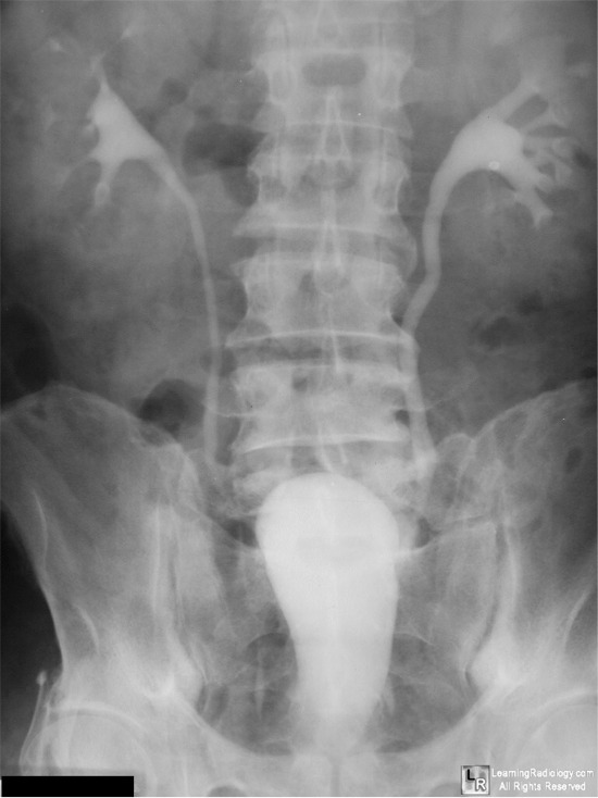
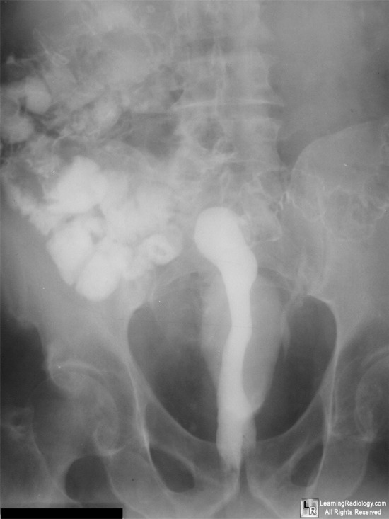
Pelvic Lipomatosis. Image from an intravenous pyelogram (IVP) at left shows bilateral compression of the urinary bladder (black arrows). The image at right, taken several hours after an upper GI series and the IVP, shows lateral compression of barium in the rectum (red arrows). There is increased lucency in the pelvis (white arrows) from excess fat deposition.
For more information, click on the link if you see this icon
For this same photo without the annotations, click here and here
Pelvic Lipomatosis eMedicine Rodriguez, J and Malik, A.
Adult and Pediatric Urology. Gillenwater, J; Grayhack, J; Howards, S and Mitchell. M. Lippincott Williams & Wilkins, 2001
|
|
|
