|
|
Chondroblastoma
Codman Tumor
General Considerations
- Rare, benign bone tumor that arises in the epiphysis of long bones in skeletally immature patients
- Usually occurs under the age of 20; males greater than females
- Most common locations are in lower extremity (70%)
- Femur most common
- Most chondroblastomas of the proximal femur occur in the greater trochanter
- Proximal tibia
- Proximal humerus
- Most occur in humeral head
- About 10% occur in the small bones of the hands and feet
- May also occur in apophyses
- Most common tumor of patella
Clinical Findings
- Pain
- Limitation of motion
- Redness
- Swelling
Imaging Findings
- Conventional radiographs are usually diagnostic
- Geographic lytic lesion arising in the epiphysis of a child
- May have scalloping or expansion of epiphyseal cortex
- May extend to adjacent metaphysis
- Eccentric
- Up to half may have internal calcifications
- Well-defined sclerotic margin
- CT or MRI are used to evaluate
- Extent of involvement of epiphysis
- Proximity of lesion to articular cartilage
- On MRI
- Low signal intensity on T1
- High or variable intensity on T2
Differential Diagnosis
- Counterpart of chondroblastoma in a child is giant cell tumor (GCT) of an adult

- GCT arise later in life (20-30)
- Frequently extend to metaphysis
Treatment
- Curettage
- Packing with allograft or autograft bone chips or methylmethacrylate
Complications
- High rate of recurrence (up to 1/3)
- Most common locations for recurrence to occur are proximal femur and pelvis
- Pathologic fractures
- Malignant transformation
- Pulmonary metastases or local invasion are possible, even with “benign” tumors
Prognosis


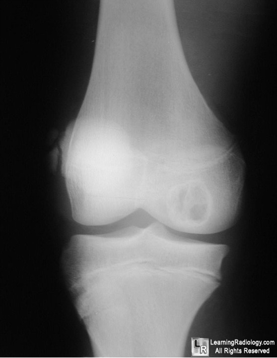
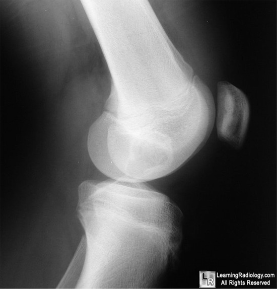
Chondroblastoma. A geographic, lytic lesion is seen in the epiphysis (white and blue arrows)
of a skeletally immature (red arrow) male's distal femur. The lesion has a sclerotic margin and is septated.
It is confined to the epiphysis. The findings are characteristic of a chondroblastoma, a rare benign bone tumor.
For more information, click on the link if you see this icon 
For this same photos without the annotations, click here and here
Wheeless Textbook of Orthopedics
eMedicine Chondroblastoma: Fines, B and Stacy, G
|
|
|
