|
|
Pyelovenous Backflow
General Considerations
- Refers to the retrograde flow of contrast material out of the intrarenal collecting system due to a small rupture of a calyceal fornix
- Almost always seen with increased pressure in the collecting system such as from
- Retrograde pyelography
- Antegrade pyelography (urogram), due to obstruction of the collecting system almost always by a ureteral calculus
- Obstruction is usually in the ureter
- Rupture decompresses collecting system
- Rarely, from vesico-ureteral reflux
Clinical Findings
- Patients with acute ureteral obstruction may report relief of symptoms when rupture of collecting system occurs
Imaging Findings
- Types of backflow include
- Pyelotubular
- Backflow into terminal collecting ducts (papillary ducts)
- Produces wedge-shaped striated area or blush extending from a calyx
- Usually considered normal phenomenon
- Pyelointerstitial
- Interstitium of kidney
- Like pyelotubular, produces wedge-shaped striated area or blush extending from a calyx, sometimes out to the edge of the renal cortex
- Pyelosinus
- Most common form
- Contrast enters the renal sinus and tracks along infundibulae, renal pelvis and proximal ureter
- Can obscure the collecting system
- May be seen with excretory urography (antegrade)
- Pyelolymphatic
- Contrast enters peripelvic lymphatics
- Multiple small channels in the hilum of kidney directed towards the para-aortic lymph node chain are visualized
- May be seen with excretory urography (antegrade)
- Pyelovenous
- Rare
- Contrast enters interlobar, arcuate renal veins
- Static radiographs rarely catch this swift-moving event but rapid sequence images may
Treatment
Complications
- Urinoma
- Retroperitoneal fibrosis

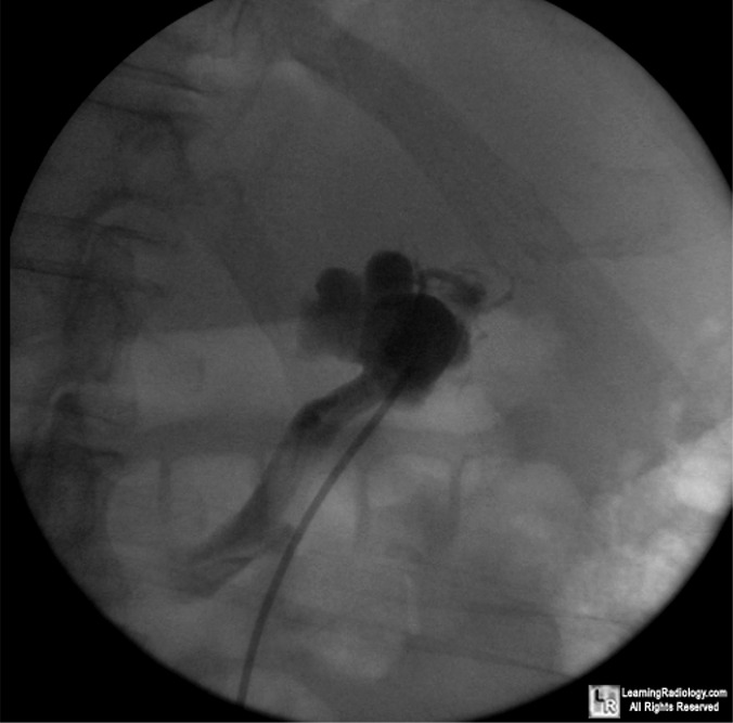
Pyelovenous Backflow. Single image from a retrograde pyelogram shows the catheter
(dotted white arrow) in the left ureter. There is a slightly dilated collecting system (dotted yellow arrow).
There is extraluminal contrast in venous structures (yellow arrow), including the left renal vein (white arrow).
For more information, click on the link if you see this icon 
For this same photo without the annotations, click here
Medcyclopedia
Pyelovenous Backflow Seen on CT Urography Nemeth A and Patel S: AJR 2004; 182:532-533
|
|
|
