|
|
Barium Enema Perforation
General Considerations
- Barium sulfate is used as a contrast agent for, amongst other things, barium enema examinations
- It is inert, radiopaque and provides excellent anatomical detail
- On very rare occasions, a perforation may occur allowing the administered barium to leak into the peritoneal cavity
- Barium peritonitis may occur from an inflammatory reaction
- Intestinal content, e.g. fecal material, which also spills into the peritoneal cavity at the same time may play a role in producing sepsis
- “Sterile” barium contamination will still cause peritonitis, but it may not be accompanied by the same degree of sepsis
- Perforations may occur
- At the enema tip
- From inflation of the retention balloon
- From a recent full-thickness biopsy of the bowel
- In diseased bowel, such as carcinoma, diverticulitis and ulcerative colitis
- Rarely, from increased hydrostatic pressure
Clinical Findings
- Signs of acute peritonitis minutes to hours after the study
- Fluid is shifted into the peritoneal cavity by the body following a perforation
- This can result in hypovolemia unless IV fluids are administered
- Abdominal pain and distension
- Rigidity
Imaging Findings
- Barium may leak freely into the peritoneal cavity, coating the surface of the bowel and liver
- Barium may also dissect into the wall
- Rarely, the perforation may be retroperitoneal
- Extraperitoneal perforations result in extra-luminal barium localized to the pelvis
Type of Contrast for Suspected Perforations |
Suspected perforation of the |
Use |
Then |
Pharynx or esophagus |
Water-soluble contrast first |
If negative, can use barium next |
If aspiration is a concern |
Barium or iso-osmolar water soluble contrast |
If negative, then usually CT |
Stomach or duodenum |
Water-soluble contrast first |
If negative, then usually CT |
Colon |
Water-soluble contrast |
If negative, then usually CT |
Differential Diagnosis
Treatment
- Laparotomy for repair of the perforation
- Removal of as much of the barium as possible
Complications
- Granulomas with adhesions
Prognosis
- Some studies have previously indicated a 50% mortality if both barium and feces were released into the peritoneal cavity
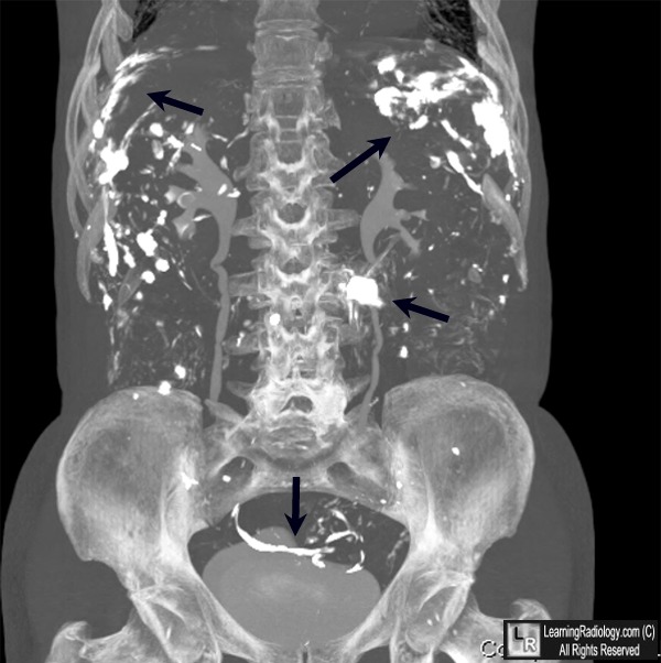
Barium in the peritoneal cavity. The black arrows point to metallic density barium
contrast that had spilled into the peritoneal cavity from a barium enema performed
elsewhere 15 years earlier. The patient was having a CT urogram
for an un-related problem. Note how the barium had flowed into the paracolic gutters
and around loops of bowel.
For more information, click on the link if you see this icon 
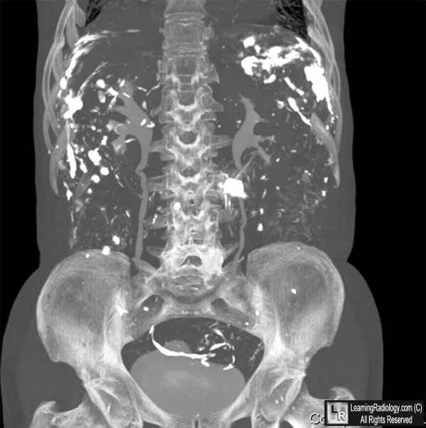
For this same photo without the arrows, click here
Williams SM, Harned RK. Recognition and prevention of barium complications. Curr Probl Diagn Radiol. 1991; 20(4):123-51.
|
|
|
