|
|
Eosinophilic Granuloma of Skull
Langerhans Histiocytosis
- Also known as eosinophilic granuloma(tosis)
- Proliferative disorder of the Langerhans cells
- Normally found in the skin (and a few other organs) and serve as antigen-presenting cells
- Rare diseases, affecting neonates up to adults
- 2:1 male to female predominance
- Prognosis
- Mortality and morbidity are associated with the clinical presentation and age of onset of the disease
- Worst prognosis for neonates presenting with the disseminated form
- Three clinical forms
- Acute disseminated langerhans cell histiocytosis (aka Letterer-Siwe disease)
- Occurs most frequently in infants 2 years of age or younger (and occasionally adults)
- Presents with multi system organ involvement
- Cutaneous lesions resembling seborrheic dermatitis involve the scalp, face, trunk and buttocks as the dominant clinical feature (nearly 80% of patient will have this)
- Infiltration of bone marrow and other organs lead to concurrent hepatosplenomegaly, lymphadenopathy, pulmonary lesions, anemia, thrombocytopenia, recurrent infections (otitis media)
- Eventually, there are destructive osteolytic bone lesions
- If untreated, this disease is rapidly fatal
- With chemotherapy, 5 year survival rate is approximately 50 percent
- Unifocal langerhans cell histiocytosis (aka Eosinophilic granuloma or granulomatosis)
- Usually only affects the skeletal system of young adults
- Typically presents as an osteolytic lesion involving the
- Calvaria
- Vertebra
- Rib
- Mandible
- Femur
- Ilium
- Scapula
- Bony lesions are usually asymptomatic
- In some cases, can cause pain and even pathologic fractures
- Pulmonary lesions may be the only presenting symptom and organ involved, especially in adults

- Skeletal lesion is usually indolent in nature
- Can heal spontaneously or be cured by local excision or irradiation
- Pulmonary lesions are typically followed and treated with supportive care
- Multifocal langerhans cell histiocytosis (aka Hand-Schuller-Christian disease)
- Triad
- Diabetes insipidus
- Exopthalmus
- Holes in the bone, usually the head (calvarium)
- Commonly affects children
- Can lead to
- Lymphadenopathy
- Hepatomegaly
- Splenomegaly
- Diabetes insipidus is secondary to infiltration of the posterior pituitary stalk by the Langerhans cell
- About a third of these patients will also display cutaneous lesions
- Some will experience spontaneous regression while others can be treated with chemotherapy
Quick Facts
Letterer-Siwe Disease
- 10% of histiocytosis X
- Acute disseminated, fulminant form
- Age at onset
- Several weeks after birth to 2 years
- Pathology
- May be confused with leukemia
- Symptoms
- Hemorrhage, purpura
- Severe anemia
- Fever
- Hepatosplenomegaly and lymphadenopathy
- Bone involvement in 50%
- Prognosis: 70% mortality rate
Hand-Schuller-Christian
- 15-40% of Histiocytosis X
- Triad of:
- Exopthalmus (33%)
- Diabetes insipidus (30-50%)
- Lytic skull lesions
- Pathology
- May simulate Ewing's sarcoma
- Age at onset
- Target organs
- Bone
- Lytic skull lesions with overlying soft tissue nodules
- Large geographic skull lesions
- "Floating teeth" with mandibular involvement
- Soft tissue
- Hepatosplenomegaly is rare
- Lymphadenopathy which may be massive
- Lung
- Cyst and bleb formation with spontaneous PTX
- Ill-defined diffuse nodular disease often leading to fibrosis and honeycombing
- Prognosis: spontaneous remissions and exacerbations
Eosinophilic granuloma
- 60-80% of Histiocytosis X
- Usually confined to bone
- Age at onset
- 5-10 years highest frequency
- Male predominance 3:2
- Location
- Calvarium>mandible>spine>ribs>long bones
- Most are monostotic (50-75%)
- Target organs
- Skull (50%)
- Diploic space of parietal bone most often
- Round or ovoid punched out lesions with beveled edge
- Sclerotic margin during healing phase
- Beveled edge=hole-within-a-hole
- Button sequestrum- bony sequestrum within lytic lesion
- Axial skeleton (25%)
- "Vertebra plana"-"coin-on-edge"(Calve disease)=collapse of vertebral body, mostly thoracic
- Most common cause of vertebra plana in children
- Proximal long bones (15%)
- Expansile, lytic lesions, mostly diaphyseal
- Soft tissue mass
- Laminated periosteal reaction
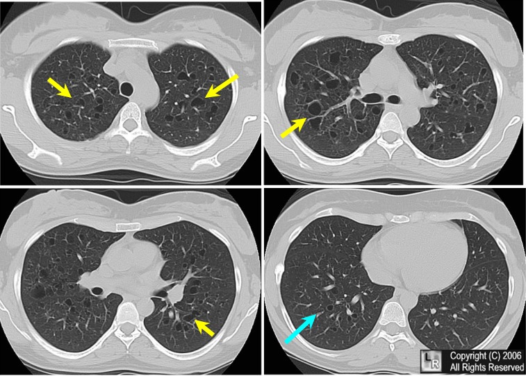
- Lung (20%)
- Age peak between 20-40 years
- Multiple small nodules
- Predilection for apices
- Prototype for honeycomb lung
- Recurrent pneumothoraces (25%)
- Rib lesions with fractures common
- Nuclear Medicine
- Negative bone scans in 35%
- Bone lesions usually not Ga-67 avid
- Ga-67 may be helpful in detecting non-osseous lesions
- Prognosis: excellent
 
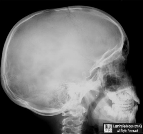
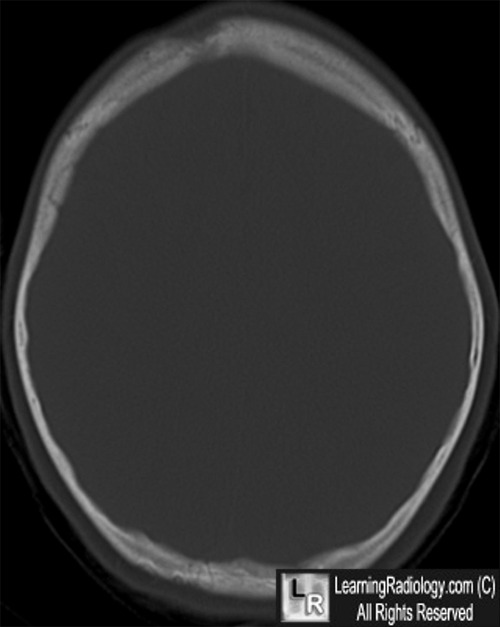
Eosinophilic granuloma of the skull. On the left, lateral skull radiograph demonstrates two lytic lesions in the frontal and parietal bones (white circles) with characteristic "beveled edges." The CT scan (at right)..shows the lesion in the right frontal bone (white arrow) and the beveling of the destructive process.
For more information, click on the link if you see this icon 
For these same photos without the annotations, click here or here
Sources: Cotran, Kumar, Collins. Pathologic Basis of Disease, 6th edition. Saunders, 1999.
EMedicine. Langerhans Cell Histiocytosis. A. Selim MD
|
|
|
