|
|
Paget's Disease of the Femur
General Considerations
- Femur is one of the bones most commonly involved by Paget’s disease

- There is accelerated bone turnover in Paget’s at active sites resulting in bones that have
- Thickened cortices, and
- Are mechanically inferior to normal bones
- Bone softening leads not only to bowing of long bones, but also
Clinical Findings
- Anterior and lateral bowing of the femur
- Warmth and tenderness of the thigh
Imaging Findings
- Characteristic changes of Paget’s disease of bone include
- Coarsening of the trabecular pattern, and
- Increased size of the bone
- The bowing in Paget’s disease of the femur is anterior and lateral and usually involves most of the femoral shaft
Differential Diagnosis
- The bowing in Paget’s disease is not the same as that seen with the true Shepherd's crook deformity which classically occurs in the polyostotic form of fibrous dysplasia and osteogenesis imperfecta
- In those conditions only the proximal femur and femoral neck are involved, producing a coxa vara deformity

Actual Shepherd’s Crooks for sale
by the Midstates Wool Growers Cooperative Association
Treatment
- If severe, osteotomy has been used to reduce the degree of bowing
Complications

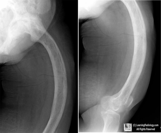
Bowing of Femur in Paget's Disease. Frontal (left) and lateral (image on right) radiographs of the left femur demonstrate anterior and lateral bowing of the femur. The trabecular pattern is coarse (white oval) and the cortices are thickened (white arrows), both characteristic of Paget's disease of bone.
For more information, click on the link if you see this icon
For this same photo without the annotations, click here
|
|
|

)