|
|
Hemangioma of the Spine
General Considerations
- Benign
- Most often located in lower thoracic, upper lumbar spine
- Skull is second most common location (spoke-wheel appearance)
- Mostly asymptomatic
- More frequent in females
- Peak incidence in 40’s
- Multiple in up to 1/3 of cases
- Most often occur in the medullary cavity of bone
- Microscopically, there is hamartomatous proliferation of vascular tissue
- Classified as to cavernous, capillary, arteriovenous and venous
- Spine hemangiomas are usually capillary type; skull are cavernous
Clinical Findings
- Usually asymptomatic
- Very slow growing
- No known malignant potential
- Over 40, patients may present with pain from compression fracture
Imaging Findings
- Conventional radiography is usually the first means of imaging hemangiomas
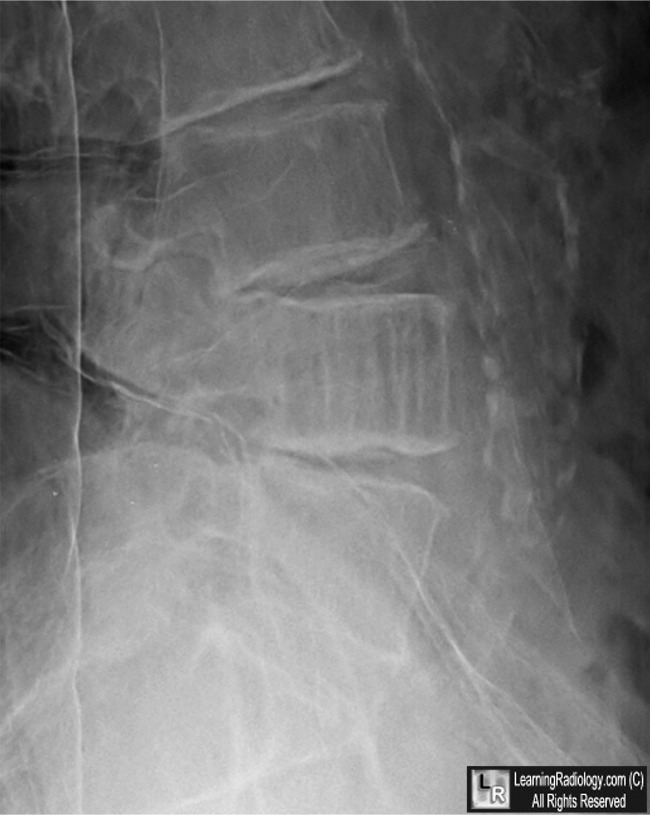
- Prominent trabecular pattern from resorption of trabeculae by enlarged vascular channels produces
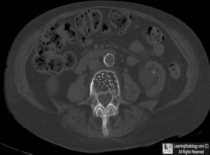
- Overall density of vertebral body is increased
- Cortex is not thickened and vertebral body is not increased in size
- Small hemangiomas will not be visible on conventional radiographs
- Corduroy (aka accordion, honeycomb, polka-dot) spine from coarse trabeculae seen in cross section
- Thickened vertebral trabeculae produce a polka-dot appearance
- Bone destruction and soft tissue extension may be present but are rare
- Allows for diagnosis of soft-tissue extension
- Increased signal intensity on both T1 (high fate content) and T2 (increased vascularity)
- Usually normal uptake on bone scan
Differential Diagnosis
- Paget disease
- Metastases
- Lymphoma
- Multiple myeloma
Treatment
- Observation
- Treatment is instituted only if they are symptomatic and may include
- Vascular embolization prior to surgery
- Surgical excision
- Vertebroplasty
- Ethanol injection
Complications
- Pathologic fracture
- Hemorrhage, when it occurs, is usually iatrogenic
- Thrombosis
- Displacement of adjacent nerves producing pain


Hemangioma of the Spine. (Top) Close-up lateral radiograph of the lower lumbar spine shows the classical coarse linear striation in the body of L4 (white circle). (Bottom) An axial CT scan image of the lower abdomen demonstrates a vertebral body with a very prominent trabecular pattern characteristic of the corduroy or polka-dot appearance of a hemangioma of the spine (yellow circle). The cortex is not thickened and the surrounding soft tissues are normal.
For these same photos without the arrows, click here and here
For more information, click on the link if you see this icon 
Hemangioma of Bone eMedicine Chasi, I and Hide, G
|
|
|
