|
|
Diffuse Axonal Injury
DAI
General Considerations
- Result of deceleration injuries, especially in high-speed motor vehicle collisions
- Also a major cause of morbidity in “Shaken Baby Syndrome”
- Most frequent cause of persistent vegetative state following trauma
- Result of rotational shear injuries that most often occur at the gray-white matter junction
- Extent of injury is usually worse than that depicted by imaging
- Shearing leads to edema, axoplasmic leakage, retraction ball formation and wallerian degeneration
- Brainstem function is typically unaffected
Clinical Findings
- Damage occurs at time of injury and then when secondary swelling occurs
- Immediate loss of consciousness
- Most, but not all, have no period of lucidity
- Brainstem functions remain intact so it is rarely a cause of death
Imaging Findings
- Diffuse
- Bilateral
- Majority of lesions (80%) are multiple
- Occur at the gray-white matter junction
- Lesions are most frequently ovoid, larger centrally than peripherally
- Frequently involved are the
- Frontal and temporal lobes
- Posterior body and splenium of the corpus callosum
- Caudate nuclei
- Thalamus
- Tegmentum
- Internal capsule
- More frequently associated with hemorrhage
- MRI is the preferred method of study but CT is usually more available
- 50-89 of patients with DAI may have a normal CT scan on presentation
- Small petechial hemorrhages located at gray-white matter junction and corpus callosum are characteristic but occur in only about 20%
- There may also be small. Focal areas of decreased attenuation secondary to edema
- Gradient-echo sequences are very useful in demonstrating paramagnetic effects of petechial hemorrhages
- Most common MRI finding
- Presence of multiple focal areas of abnormally bright signal on T2-weighted images in the white matter of the temporal or parietal corticomedullary junction or in splenium of corpus callosum.
- Hemorrhagic lesions appear hyperintense on T1-weighted images
- Non-hemorrhagic lesions appear hyperintense on T2-weighted sequences
- Gradient-echo sequences are very useful in demonstrating paramagnetic effects of petechial hemorrhages
Differential Diagnosis
- Multiple sclerosis
- Embolic or hemorrhagic stroke
Prognosis
- Over 90% remain in persistent vegetative state
- Chances of this occurring are greater with lesions that are supratentorial, involve the corpus callosum or corona radiata
- Prognosis worsens with multiplicity of lesions

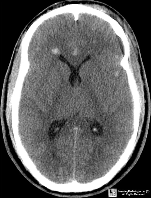
Diffuse Axonal Injury. An axial, non-enhanced CT image of the brain demonstrates multiple small petechial hemorrhages at the gary-white matter junction, the caudate nucleus and the corpus callosum, characteristic of diffuse axonal injury in this male who was in a motor vehicle accident.
For more information, click on the link if you see this icon 
For this same photo without the annotations, click here
Diffuse Axonal Injury eMedicine Wasserman, J; Koenigsberg, R
|
|
|
