Learning Radiology xray montage


Free Intraperitoneal Air
Pneumoperitoneum


Causes of free air

  • Disruption of wall of hollow viscus

    • Blunt or penetrating trauma

    • Perforating foreign body

    • Iatrogenic perforation

      • Laparoscopy / laparotomy

        • Absorbed in <7 days depending on initial amount of air introduced and body habitus

      • Leaking surgical anastomosis

      • Endoscopic perforation

      • Diagnostic pneumoperitoneum

    • Diseases of GI tract

      • Perforated gastric or duodenal ulcer

      • Ingested foreign-body perforation

      • Diverticulitis (though abscess is usually walled-off)

      • Necrotizing enterocolitis with perforation

      • Inflammatory bowel disease (e.g. toxic megacolon)

      • Perforated appendix rarely causes a large amount of free air because appendiceal obstruction leaves only a few cc’s of air in appendix if it perforates

      • Obstruction from neoplasm, imperforate anus, Hirschsprung’s disease, meconium ileus

  • Through peritoneal surface

    • Abdominal needle biopsy or catheter placement

    • Mistaken thoracentesis or chest tube placement

    • Endoscopic biopsy

  • Extension from:

    • Dissection from pneumomediastinum

    • Bronchopleural fistula

    • Rupture of urinary bladder

    • Penetrating abdominal injury

  • Intraperitoneal

    • Gas forming peritonitis

    • Rupture of abscess

Imaging findings

  • Best seen with horizontal x-ray beam

  • Three major findings
    • Free air beneath diaphragm (crescent sign)

    • Visualization of both sides of the bowel wall

      • "Rigler’s sign"

        • Normally, only the inside of the bowel (the lumen) is visible unless free air outlines the outer surface of the wall

    • Visualization of the falciform ligament


      • Long vertical line to the right of midline extending from ligamentum teres notch to umbilicus; most common structure outlined

      • "Football sign" = large pneumoperitoneum outlining entire abdominal cavity with “laces” representing falciform ligament

  • RUQ gas (best place to look for small collections)

    • Single large area of hyperlucency over the liver

    • Oblique linear area of hyperlucency outlining the posteroinferior margin of liver

    • Triangular area of free air trapped below central tendon of diaphragm

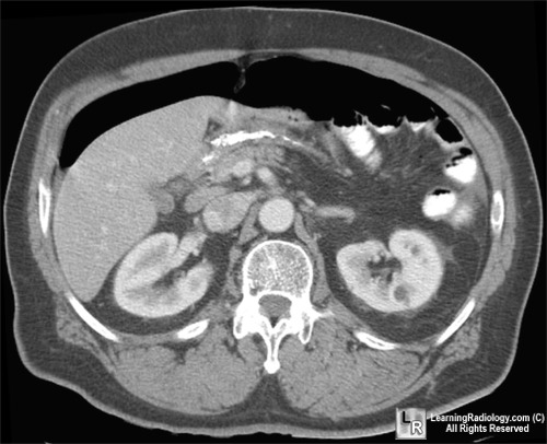
Free air. There is a large quantity of free air in this patient's abdomen. The image is obtained with the patient supine. Free air has risen above the liver and bowel (red arrows). The air is not contained within any visible bowel wall. The falciform ligament is surrounded by air on either side (white arrow).
For additional information about this disease, click on this icon above.
For this same photo without the arrows, click here