|
|
Unicameral Bone Cyst
Simple Bone Cyst, Solitary Bone Cyst
General Considerations
- Common (3-5% of primary bone tumors)
- Benign
- Solitary lesion
- Most common in proximal humerus and femur
- Also Iliac bone and calcaneous, especially over age 20
- Most common between 4-10 years old
- Filled with clear, yellowish fluid
- May contain giant cells and hemosiderin
Clinical Findings
- Asymptomatic
- If fractured, then
Imaging Findings
- Conventional radiography is the study of choice
- Most common in proximal humerus (in patients under 20) and femur in skeletally immature male
- Solitary, lytic metaphyseal lesion adjacent to, but not crossing, the epiphyseal plate
- Migrates towards diaphysis during growth of child
- Well-defined margins with narrow transition zone
- May be slightly expansile
- Long axis of lesion is parallel to long axis of bone
- May have thin sclerotic margin
- Endosteal scalloping and erosion
- Fallen Fragment Sign represents a fragment of bone that falls into the cyst and then into a dependent position
- Although uncommon, it is pathognomonic for a simple cyst because it indicates the fluid nature of the interior of this lytic lesion
- On CT, they may be shown to contain air-fluid or fluid-fluid levels
- On MRI. They will have low signal intensity on T1 and high on T2
- Lesions which have fractured will have a heterogeneous signal on T1 and T2 because of the hemosiderin
- Their periphery may enhance with Gadolinium
- Photopenic on bone scan
Differential Diagnosis
- Fibrous dysplasia
- Ground-glass; more irregularly shaped
- Eosinophilic granuloma
- Look for other lesions; beveled-edge; vertebra plana
- Chondroblastoma
- Chondromyxoid fibroma
- More expansile and eccentric; more rare
- Brown tumor
- Other signs of hyperparathyroidism
- Aneurysmal bone cyst
- Enchondroma
- Smaller bones; internal calcifications
Treatment
- Curettage and bone grafting, nailing, injection of bone marrow or cryotherapy to prevent pathologic fracture
- Methylprednisolone injections have been used to promote healing
Complications
- Pathologic fractures in 50-65%
- Growth arrest in affected limb
Prognosis
- Usually undergo spontaneous regression

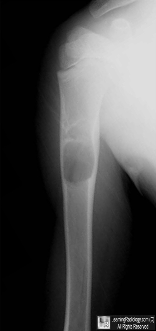
Unicameral Bone Cyst. A single frontal radiograph of the shoulder
demonstrates a geographic lytic lesion (yellow arrow) in the metadiaphysis
of the humerus with a sharp zone of transition and inner septations. There is no pathologic fracture through the lesion.
For this same photo without the arrow, click here
For more information, click on the link if you see this icon 
Simple Bone Cyst eMedicine Eu-Leong Harvey Teo and Wilfred CG Peh.
|
|
|
