|
|
Uterine Fibroids
General Considerations
- Uterine fibroids are leiomyomas, benign tumors of smooth muscle origin
- Occur in between 20-50% of women > 30 years of age
- Fibroids may enlarge with elevated estrogen levels
- They enlarge during the first trimester of pregnancy
- Uterine fibroids diminish in size after menopause
- More common in blacks than whites (3:1)
- Most are intramural, i.e. middle of myometrium
- Most fibroids are in the fundus and body of the uterus
- Others can be subserosal or exophytic, or
- Submucosal and subendometrial (rarely)
Clinical Findings
- Most women are asymptomatic
- Symptoms can include
- Menorrhagia (increased duration or flow)
- Frequently from a submucosal fibroid
- Pain
- Urinary frequency, urgency or incontinence
- Infertility
Imaging Findings
- Ultrasound is the study of choice
- Conventional radiography
- Soft tissue mass arising from the pelvis but separate from the urinary bladder
- Amorphous, flocculent calcifications in the pelvis
- Displacement of bowel gas up and out of the pelvis
- Ultrasound
- Echogenic mass if fibrosis prevails
- Hypoechoic, solid mass if muscle component is prevalent
- Sharp discrete shadows
- Anechoic features if central portion of fibroid has degenerated
- MRI
- Low/intermediate signal intensity of T1 and T2 weighted images
- High central signal intensity on T2 from hemorrhage
- May have hyperintense rim
- With contrast, most are hypointense, about 25% isointense and 10% hyperintense to myometrium
- CT
- Mass containing mixed densities, low attenuation if necrotic and higher attenuation if calcified or hemorrhagic
Differential Diagnosis
- Ovarian neoplasm or cyst
- Dilated urinary bladder
- Intrauterine pregnancy
- Hydatidiform mole
Treatment
- Surgery or arterial embolization for pain and menorrhagia
Complications
- Rarely (< 1%), malignant degeneration to a leiomyosarcoma
- Exophytic fibroid can torse and cause pain
- Infertility from interference with implantation
- During pregnancy
- Intrauterine growth retardation
- Postpartum hemorrhage
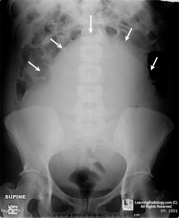 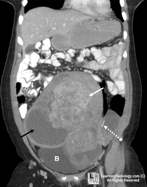
Uterine Fibroids. Conventional radiograph on the left shows displacement of the bowel gas out of the pelvis because of a large soft tissue mass (white arrows) arising from the pelvis and extending into the lower abdomen. Contrast-enhanced coronal reformatted CT scan of the abdomen on right demonstrates a large pelvic mass comprised of degenerating fibroids (black arrow), hemorrhage (white arrow) and calcification (dotted white arrow).
For same photos without annotations, click here or here
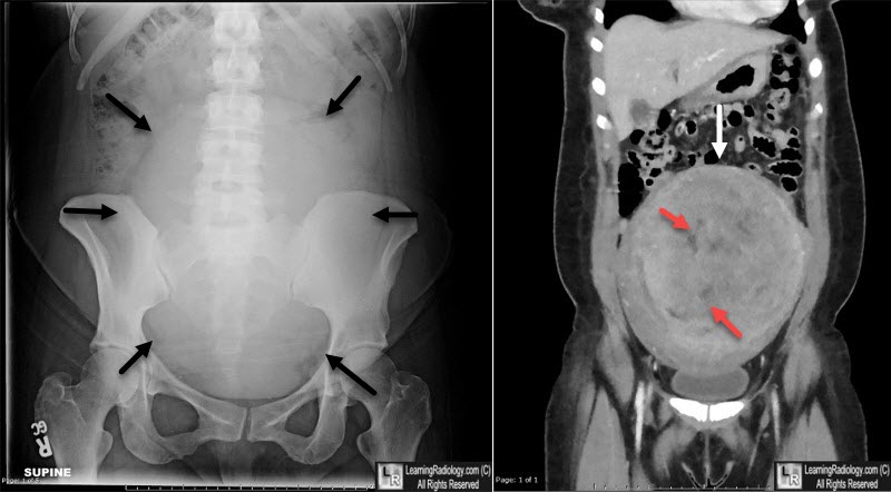
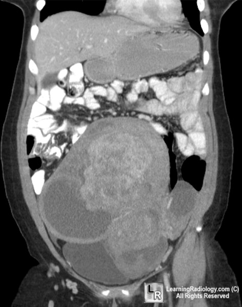
Uterine Fibroids. Conventional radiograph on the left shows a large soft tissue mass (black arrows) arising from the pelvis and extending into the lower abdomen. Non-contrast-enhanced coronal reformatted CT scan of the abdomen on right demonstrates a large pelvic mass comprised of a large fibroid (white arrow), with some areas of degeneration (red arrows).
For additional information about this disease, click on this icon if seen above.
Williams Obstetrics 22nd Ed McGraw Hill
|
|
|

)
)