|
|
Atelectasis of the Left Lower Lobe
General Considerations
- All types of atelectasis involve loss of volume in some or
all of a lung with resultant increased density of the involved lung
- The atelectasis referred to here is that caused by bronchial obstruction, usually a tumor (i.e. a bronchogenic carcinoma), a foreign body or a mucus plug
Signs of atelectasis
Lower Lobe Atelectasis
- Lower lobe is usually tethered to the hemidiaphragm and mediastinum by the inferior pulmonary ligament (IPL)
- The IPL attaches the lower lobe to the mediastinum
and may be seen normally on CT scans of the chest
- The inferior pulmonary ligament is actually not a
ligament but reflections of the pleura (visceral and parietal) inferior to the hilum extending downward to the
diaphragmatic pleura
- It is on the medial border of the left lower lobe
- There is no superior pulmonary ligament as a
counterpart
- With collapse, there is rotation of the major fissure
posteriorly and medially
Imaging signs of Left Lower Lobe Atelectasis
- Wedge-like density behind heart with base of triangle on the left hemidiaphragm and the apex at the hilum on frontal view
- Silhouetting of all or just the medial portion of the left hemidiaphragm
- Posterior displacement of the major fissure on the lateral
view 
- Hilum may be displaced inferiorly
- Descending branch of the pulmonary artery may disappear
- There may be a shift of the heart towards the left which
means less of the right heart border or none of the right heart
border may project to the right of the spine
- There may be a a spine sign due to superimposition of
the non-aerated lower lobe and the spine on the
lateral view
- In severe atelectasis of the lower lobe, the atelectatic lobe
is so small it may not be recognizable on a conventional
radiograph
- The hemidiaphragm may be seen throughout its
length. i.e. there is no silhouette sign
- The normal contour at the top left of the aortic knob may become invisible
- Appearance of left lower lobe atelectasis is similar to right
lower lobe atelectasis
Associations
- Following cardiac surgery, especially in which there is topical hypothermia of the heart and phrenic nerve
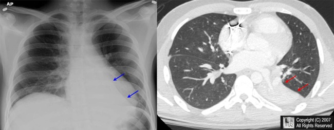
Left lower lobe atelectasis. The blue arrows point to the edge of a triangular region of increased density
in the left cardiophrenic sulcus representing the major fissure with the atelectatic left lower
lobe medial to it. The red arrows on the axial CT of another patient show the atelectatic left lower
lobe bounded by the displaced major fissure.
For this same photo without arrows, click here
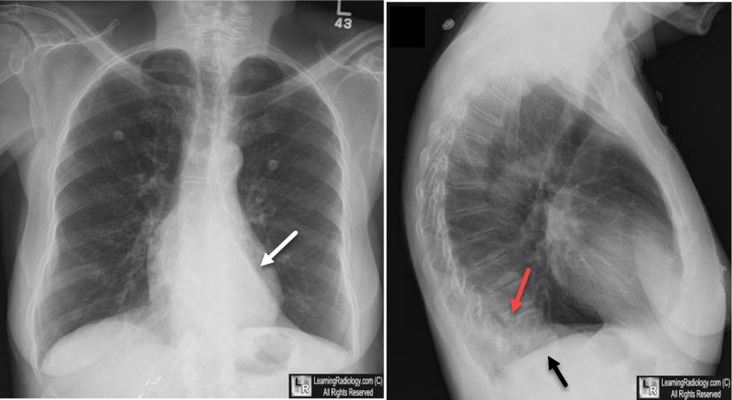
Left lower lobe atelectasis. The frontal view demonstrates a sharp edge which is the major fissure and a triangular density at the left base behind the heart (white arrow). On the lateral view, the area of increased density is seen posteriorly (red arrow) while only the right hemidiaphragm is visualized since the left is being silhouetted by the atelectasis.
For additional information about this disease, click on this icon if seen above.
British Journal of Radiology 74 (2001),89-97 © 2001 Lobar atelectasis: diagnostic pitfalls on chest radiography K Ashizawa, MD, K Hayashi, MD, N Aso, MD and K Minami, MD
Radiology, Vol 148, 479-483, Copyright © 1983 by Radiological Society of North America Inferior pulmonary ligament: computed tomographic appearance RC Rost Jr and AV Proto
|
|
|

)
)
)