|
|
Talar Beak
Tarsal Coalition
General Considerations
- Rare (1% incidence)
- Fusion of two or more tarsal bones in the hindfoot (talus and calcaneous) or midfoot (cuboid, navicular and 3 cuneiforms)
- Most common coalitions (90% of total) are
- Calcaneous and navicular (calcaneonavicular)
- Calcaneous and talus (talonavicular)
- Fusion may be congenital or acquired
- Congenital form is familial
- Autosomal dominant with near complete penetrance
- Bilateral 50% of time
- More common in males than females
- Acquired form may be due to trauma, infection, arthritis, surgery
Types
- Complete coalition
- Incomplete coalition
- Cartilaginous (synchondrosis)
Clinical Findings
- Most are asymptomatic
- When symptomatic, symptoms begin in 2nd decade of life
- Pain is most common symptom
- Made worse by physical activity
- Restricted range of motion
- Muscles spasms
Imaging Findings
- Conventional radiography of the foot is the study of first choice
- Calcaneonavicular fusions should all be evident on a 45° internal oblique view
- Bridge is usually from anterolateral aspect of calcaneous to dorsolateral aspect of navicular
- With fibrous and cartilaginous coalition, the joints may be narrowed, sclerotic and irregular
- Talocalcaneal coalition may be more difficult to see
- Most often involves junction between the middle facet of the talus and the sustentaculum talus
- Talar beak presumably occurs because of limitation of motion in subtalar joint
- At insertion of talonavicular ligament, periosteal reaction develops
- Beak not present in all patients with talocalcaneal coalition
- Pes planus deformity is common
- Hypoplasia of the sustentaculum tali may occur
- MRI is most sensitive to fibrous and cartilaginous fusion
Treatment
- Conservative treatment is utilized first
- NSAID (non-steroidal anti-inflammatory drugs)
- Steroid injections
- Orthotics
- Surgical interventions most commonly involve resection of the bridge and placement of fat, muscle or tendon in place of the coalition
- Arthrodesis is sometimes used with severe coalitions
Complications
- Cause of spastic peroneal flatfoot
- Bony bridge can fracture
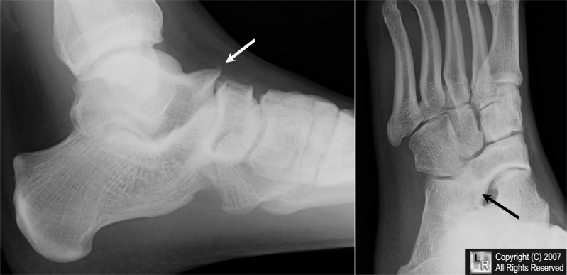
Talar Beak and Tarsal Coalition. The white arrow points to a "talar beak" seen on the dorsum of the talus
which should raise suspicion for the presence of a tarsal coalition. The oblique view of the same foot
demonstrates bony bridging between the tarsal navicular and the calcaneous.
For additional information about this disease, click on this icon if seen above.
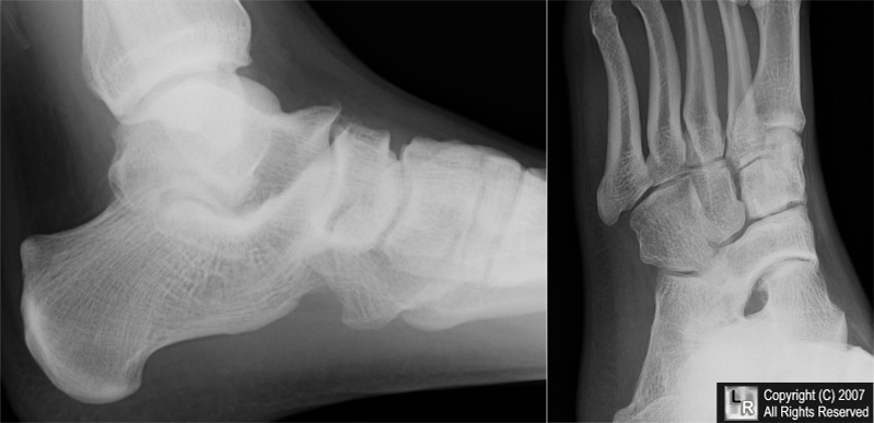
For this same photo without arrows, click here
Tarsal Coalition eMedicine Eric A Wang, MD, Amilcare Gentili, MD, Sulabha Masih, MD, and Matthew C Wang, BS
|
|
|
