|
|
Non-Union Tibial Fracture
General Considerations
- Delayed union is the term applied when a fracture has not healed within the period of time that would be considered adequate for bone healing for that particular sites
- Holds out the promise that union will eventually occur without further intervention
- For tibial fractures, healing should occur in 16 weeks
- Non-union is the term applied to a fracture that will not unite without additional intervention
- Usually by 6-9 months for tibial fractures
- Non-union of tibia is fairly common
- Estimated to range from 2-10% of all tibial fractures
Associations
- Most closely associated with the type of tibial fracture
- Open or compound fracture
- Degree of comminution
- Less soft tissue covers fracture
- High energy fractures (automobile and motorcycle accidents)
- Cigarette smoking places patient at higher risk
- Use of nonsteroidal anti-inflammatory medications may inhibit bone healing
Types
- Hypertrophic nonunions
- Exuberant callus formation
- Because of their vascularity, they have excellent healing potential
- Result from inadequate immobilization of the fracture
- Atrophic (oligotrophic) nonunions
- Absence of callus and bone ends that may be tapered and osteopenic or sclerotic
- Because of their lack of vascularity, they have poor healing potential
- A subcategory of this type may be freely movable and form a pseudarthrosis
- Normotrophic nonunions
- Share characteristics of both of the above
Imaging Findings
- Conventional radiographs are the study of first choice
- In established non-union, the ends of the fracture fragments are sclerotic and typically smooth
- Bones are joined by fibrous tissue
- CT may be helpful in establishing presence of bony bridging
- MRI is most sensitive for osteomyelitis
Differential Diagnosis
- Congenital pseudarthrosis of the tibia
- Associated with neurofibromatosis and fibrous dysplasia
Treatment
- Hypertrophic nonunions are treated with rigid stabilization
- Atrophic nonunions require augmentation
- May be in the form of bone grafting or biologic stimulation
Complications
- Infection of a previously aseptic non-union
- Malalignment
- Shortening
- Continued non-union
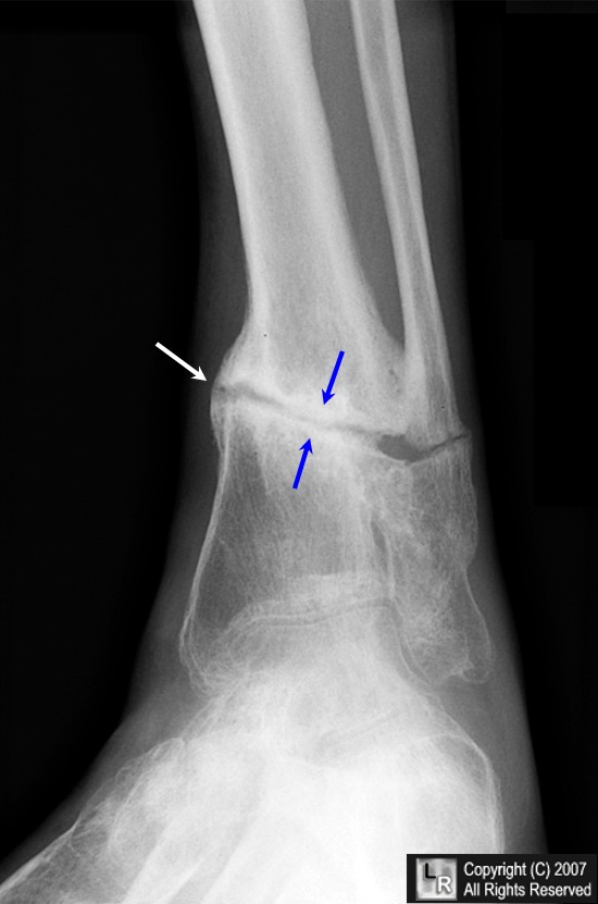
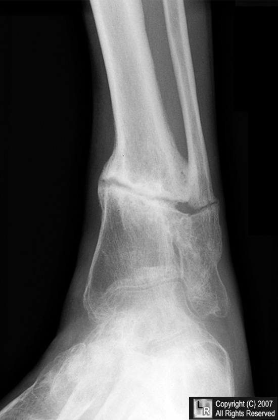
Tibial non-union. Frontal radiograph of the distal tibia shows a smooth and sclerotic line at the fracture ends (blue arrows) in a patient 14 months after the original fracture, signs of non-union. There is some external callus formation present (white arrow). There is also non-union of an associated fibular fracture.
For additional information about this disease, click on this icon if seen above.
For this same photo, click here
eMedicine Minoo Patel, MBBS, MS, FRACS, James McCarthy, MD, John Herzenberg, MD, FRCSC
Wheeless’ online textbook of Orthopedics
|
|
|
