|
|
Blowout Fracture of the Orbit
General Considerations
- Isolated fractures, most commonly of the orbital floor
- The cause is sudden, direct, blunt trauma in the form of a blow to the orbit with increase in intraorbital pressure
- The orbital rim is relatively strong so force is transmitted to the weakest parts of the orbit which “blow-out”
- Orbital floor which is the superior boundary of the maxillary sinus, or
- Medial wall (the thin lamina papyracea) which is the lateral boundary of the ethmoid sinus
- The nasal bone is also frequently fractured
- The cause is usually a large object such as baseball, fist, automobile accidents, tennis ball and kick
Clinical Findings
- Pain and tenderness
- Diplopia on upward gaze
- Due to entrapment of the inferior rectus and sometimes the inferior oblique muscles
- Enophthalmos
- Usually following initial swelling and proptosis
- Patient reports feeling of pressure in orbit when attempting to blow nose
- Facial anesthesia due to entrapment of the infraorbital nerve
- Epistaxis
Imaging Findings
- CT of the facial bones is the imaging study of choice
- Orbital emphysema
- Fracture of the floor or medial wall of the orbit
- Depression of the fracture fragment(s)
- Soft-tissue mass extending into the maxillary sinus
- Complete or partial opacification of the ipsilateral maxillary sinus from hemorrhage or edema
Treatment and complications
- Requirement for, timing of and method used for reconstruction of orbital floor is controversial
- Surgical repair, when performed, usually occurs after swelling has subsided
- If the diplopia does not resolve spontaneously
- Severe enophthalmos (>2mm)
- Large fractures (50% or more of floor)
- Consists usually of resection of periosteum and repair of hole using either bone graft, plate or synthetic material such as Teflon
- Long-standing entrapment can lead to vision impairment and enophthalmos

Blowout fracture of the orbit . Reformatted coronal CT of the facial bones demonstrates a fracture of the floor of the left orbit (white arrow) associated with orbital emphysema (blue arrow). A portion of the inferior rectus muscle (solid red arrow) projects into the maxillary sinus below (see normal opposite side--broken red arrow). There is blood in the maxillary sinus (white arrow).
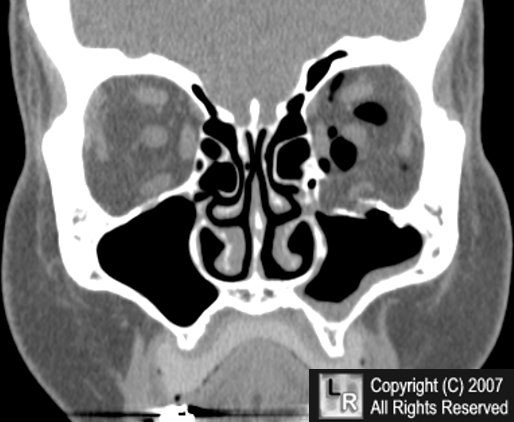
For this same photo without the arrows, click here
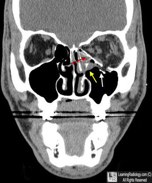
Blowout fracture of the orbit . Reformatted coronal CT of the facial bones demonstrates a fracture of the floor of the left orbit (red arrow) with a depressed fragment protruding into the left maxillary sinus (white arrow). A portion of the inferior rectus muscle (yellow arrow) projects into the maxillary sinus below.
For more information, click on the link if you see this icon 
|
|
|
