|
|
Appendicolith with Appendicitis
General considerations
- Also known as a fecolith, fecalith, coprolith
- Calcified deposit within the appendix
- Present in approximately 30% of children with acute appendicitis
- May be an incidental finding on an abdominal radiograph done for other purposes
- But, when associated with abdominal pain, there is a 90% probability of acute appendicitis
- Also 50% higher risk of appendiceal perforation
- Of some controversy, the finding of an appendicolith may be sufficient evidence to perform a prophylactic appendectomy in asymptomatic patients given the higher rate of perforation at the time of acute appendicitis
- One of several causes of obstruction of the appendiceal lumen leading to acute appendicitis which also include
- Lymphoid hyperplasia
- Foreign bodies
- Stricture
- Tumor
- Crohn’s disease
- For more on clinical and imaging findings of acute appendicitis, click here

Imaging Findings
- The role of imaging is to confirm clinically suspected appendicitis, rule out another diagnosis or a complication of the disease
- Conventional radiography (abnormalities seen in <50%)
- Plain-film findings become more distinctive after perforation, while clinical findings subside
- Calcified, frequently laminated, appendicolith in RLQ (in 7-15%)
- It may lie higher in a retrocecal appendix
- Appendicolith in acute appendicitis means a high probability for perforation (50%), especially in children
- Appendicolith is usually 1 cm in size of larger
- Other signs
- Localized dilatation of cecum from focal paralysis (cecal ileus)
- Small bowel obstruction pattern
- Soft-tissue mass and paucity or absence of intestinal gas in RLQ (more often with perforation and abscess)
- Extraluminal gas bubbles (usually only with perforation and abscess)
- Large pneumoperitoneum is rare because etiology of appendicitis involves obstruction of a very small lumen containing little air
- Focal increase in thickness of lateral abdominal wall
- Loss of properitoneal fat line on right side
- CT
- The imaging study of choice (>95% accurate) is CT, or US
- Signs of acute appendicitis include
- Distended lumen
- Circumferentially thickened and enhancing wall
- Appendicolith – homogeneous or laminated calcification in up to 25% of cases
- Peri-appendicular inflammation-linear streaky densities in peri-appendicular fat
- Peri-cecal soft-tissue mass
- Abscess
- Poorly encapsulated
- Single or multiple fluid collection(s) with air
- Extraluminal contrast material
- Focal cecal wall thickening
- "Arrowhead" sign = funnel of contrast medium in cecum centering about occluded orifice of appendix
Complications
Treatment


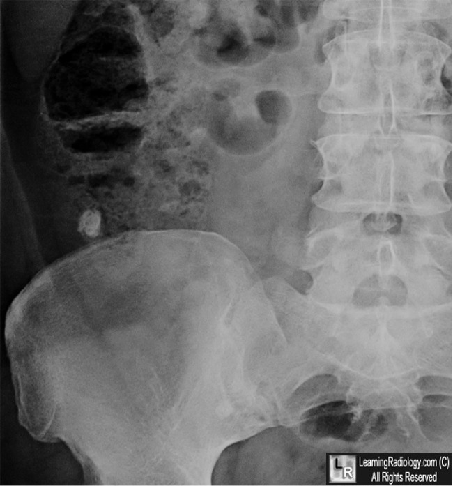
Appendicolith with Appendicitis. Upper: Frontal close-up of right lower quadrant show a laminated
stone in the region of the appendix consistent with a calcification that has formed in a viscous (white arrow).
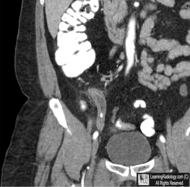
Lower: Coronal reconstruction with close-up of right lower quadrant shows a dilated appendix
with a thickened wall and surrounding infiltration of the fate (yellow arrow) containing an appendicolith (red arrow).
For these same photos, click here and here
For more information, click on the link if you see this icon
|
|
|
