|
|
Lunate Dislocation
Also Perilunate Dislocation
- General Considerations
- Mechanism of injury is usually fall onto an outstretched hand with wrist in ulnar deviation and dorsiflexion
- Wrist dislocations are uncommon but lunate is the most common bone in the wrist to dislocate
- Most severe of carpal instabilities
- Involves all the intercarpal joints and disruption of most of the major carpal ligaments
- Capitate and all other carpal bones will lie posterior to lunate on lateral radiograph
- Clinical Findings
- Wrist pain, tenderness and swelling
- Imaging findings
- Normal anatomy
- Dislocation
- Triangular appearance of lunate on frontal projection
- Not always reliable because of differences in projection
- Produces volar (anterior) dislocation and forward rotation of lunate
- Concave distal surface of lunate comes to face anteriorly
- The lunate cup “points” towards the palm
- Capitate may drop into the space vacated by lunate
- Complications
- Vascular complications may occur but are uncommon unless there is an associated fracture present, especially of the distal radius
- Soft tissue complications may include disruption of carpal ligaments which can lead to long-term carpal instability
- Many develop chronic wrist pain
Perilunate dislocation
- Triquetral and scaphoid malrotation
- Result of a fall on an outstretched, hyperextended hand
- Relatively rare
- Occurs when the lunate maintains normal position with respect to the distal radius while all other carpal bones are dislocated posteriorly
- Very commonly associated with a trans-scaphoid waist fracture
- Sometimes ulnar styloid as well
- Lunate appears triangular in shape on frontal view
- Lunate rotates forward slightly on lateral view
- On lateral view, all other carpal bones are dislocated posterior with
respect to lunate 
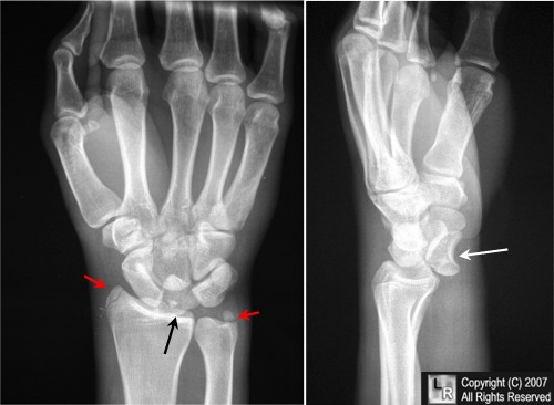
Lunate dislocation. In the frontal projection on the left, the lunate is triangular, rather than quadrilateral,
in shape (black arrow). On the lateral projection on the right, the lunate is displaced anteriorly (white arrow) and its
concave surface is directed anteriorly (toward the palm) instead of superiorly (towards the finger tips).
there are fractures of the radial and ulnar styloid (red arrows).
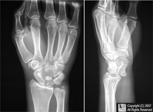
For this same photo without the arrows, click here
|
|
|

)
)
)