|
|
Metastatic Calcification
- General considerations
- Metastatic calcification refers to deposition of calcium salts in previously normal tissue
- Due to disturbance in calcium/phosphorus metabolism
- Most likely to develop when the calcium/phosphorous product exceeds 70, but may occur at normal values
- Most commonly related to persistently elevated calcium levels as in
- Primary hyperparathyroidism
- Chronic renal disease
- Seen in 60-80% of autopsied hemodialysis patients
- Milk-alkali syndrome
- Hypervitaminosis D
- Widespread bone destruction from metastases or myeloma
- Alkaline tissue environment thought to make such deposits more likely, so that most common organs involved are
- Lungs
- Gastric mucosa
- Kidneys
- Also can deposit in
- Muscle
- Subcutaneous tissue
- Heart
- Thyroid
- Liver
- Spleen
- Pancreas
- Dystrophic calcification is deposition of calcium salts in previously damaged tissue with normal calcium metabolism
- In the lung, such calcification might be seen in TB, pneumoconiosis, and sarcoid amongst many others
- Clinical findings
- Usually asymptomatic from the calcification itself
- If there is widespread deposit in the lungs, some may develop severe restrictive lung disease
- Imaging findings in the chest
- Patchy areas of consolidation
- Although calcium deposition occurs only in the interstitium, it may resemble and be confused with airspace disease on conventional radiography
- Usually affects the upper lobes
- Possibly due to high ventilation/perfusion ratio leading to high oxygen, low carbon dioxide levels and relative alkalinity
- Small, calcified nodules may be seen on CT
- May be unilateral or diffuse
- Calcification may be seen in the vessels and chest wall
- Also seen are areas of ground-glass attenuation
- Bone scintigraphy will almost always show uptake of radiotracer in the lungs
- May be more sensitive than CT
- If calcium deposit is fully mature, the bone scan agent may not be taken up in the lungs
- Prognosis
- May remain stable for years or be rapidly progressive
- Differentiation from calciphylaxis
- While also associated with chronic renal failure or renal transplantation, calciphylaxis is a disease of unknown etiology that produces necrotic skin lesions and subcutaneous blood vessel calcification
- The disease frequently starts on the lower extremities
- It frequently progresses rapidly and has a high mortality (60-80%)
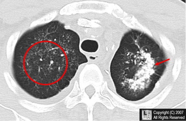
Metastatic calcification in the lungs. The red circle and red arrow point to deposits of calcium in the interstitial
tissues of both upper lobes (worse on the left with the arrow) in an appearance typical for metastatic calcification to the lungs,
seen most frequently in patients with chronically elevated calcium levels
.
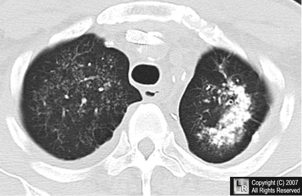
For the same photo without the arrows, click here
J HK Coll Radiol 2002;5:186-189 Metastatic Calcification Tam KF, Wang K, Fan WC
Eur Respir J 1997; 10: 1925–1927 Metastatic pulmonary calcification after renal transplantation Murris-Espin M, et al
|
|
|
