|
|
Heterotopic Ossification
HO
General considerations
- Defined as the abnormal formation of true bone within extra-skeletal soft tissues
- More common in males, especially following spinal cord injury, it is rare in young children
- Formerly called myositis ossificans
- That term has fallen out of favor because the condition is not always inflammatory and ossification occurs in soft tissues other than muscle
- Strong association exists between HO and spinal cord or traumatic brain injury
- About 20-30% of patients with neurologic deficits will develop HO, possibly higher with spinal cord injuries
- It is also seen in burn patients, following surgery, and following blunt trauma such as horse riders may develop in the adductor muscles of the leg
- There is an increased risk for HO in patients with Diffuse Idiopathic Skeletal Hyperostosis (DISH) and Paget’s Disease
Pathophysiology
- Bone morphogenetic proteins may stimulate primitive stem cells in soft tissues to form osteoblasts under certain conditions
- Following trauma, cartilage begins developing in soft tissues by 2nd week, with trabeculated bone appearing by 2-5 weeks
Clinical findings
- May cause pain and a palpable mass
- Contributes to further restricting range of motion
- Can lead to breakdown of the skin in spinal cord patients
- Post-surgical HO most commonly occurs at hip following arthroplasty which is also the most common site of HO in patients with brain or spinal cord injury
- Shoulders and elbows follow in frequency in brain injury
- Knees are uncommonly affected in brain injury but frequently affected in spinal cord injury
Imaging findings
- Conventional radiography is the study of first choice
- Nuclear bone scan is most sensitive for early detection
- Bone can be detected on conventional radiographs as early as two weeks after injury, the ossification typically starting at the periphery as a cloud-like increase in density
- Biopsy of the lesion could lead to a false-positive diagnosis of osteosarcoma unless the clinical findings are taken into account and time is allowed for the lesion to mature
- CT may show a soft tissue mass early, followed by visualization of bone earlier than can be seen with conventional radiographs
- MRI is typically not used
- Ultrasound may show abnormalities in the muscle in advance of visible ossification
- The standard for early HO detection is the triple-phase bone scan using Tc 99M MDP
- Bone scans may be positive 2-6 weeks earlier than ossification is visible
- Early in the course, only the blood pool images may be positive whereas abnormal uptake during the soft tissue phase is diagnostic later in the course of the disease
Treatment
- Prophylactic treatment may include radiation therapy using an external beam, non-steroidal inflammatory agents and oral etidronate
- Surgical resection, when performed, is usually done only after the lesion has matured, the progress of which can be monitored by bone scan
- Recurrence is relatively common following resection

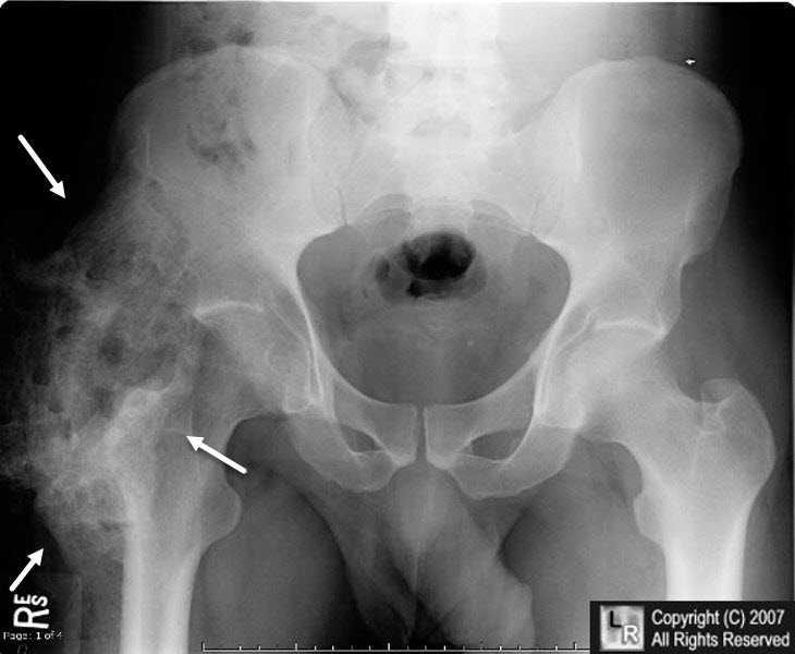
Heterotopic ossification. White arrows point to ossification (with trabeculae and cortex)
surrounding both hip joints in a young patient with a traumatic brain injury several months earlier.
For this same photo without the arrows, click here
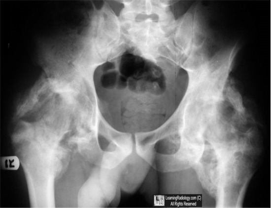
Heterotopic ossification. White arrows point to ossification (with trabeculae and cortex)
surrounding the right hip joint in another young patient with a traumatic spinal cord injury several months earlier.
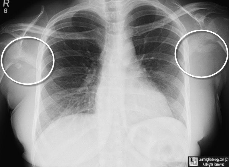
Heterotopic ossification. White circles highlight ossification in both axillae in a quadreplegic patient.
For more information, click on the link if you see this icon 
eMedicine Heterotopic Ossification Daniel S Moore, MD and Gina Cho, MD
|
|
|
