|
|
Left Superior Intercostal Vein
Aortic Nipple
- Seen as a small soft-tissue density adjacent to the lateral border of the aortic knob on a frontal radiograph in up to 10% of normal patients
- Normally the left superior intercostal vein drains the left 2nd, 3rd, and 4th posterior intercostal veins and connects the left brachiocephalic vein and the accessory hemiazygos vein
- As such, the left superior intercostal vein provides a collateral path of blood back to the heart
- The size of the left superior intercostal vein is inversely related to the size of the accessory hemiazygos vein: the smaller (or absent) the accessory hemiazygos, the larger will be the left superior intercostal vein
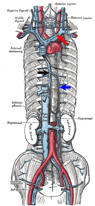
- The accessory hemiazygos vein (orange arrow below) drains the posterior intercostal veins from 3-4 intercostal spaces between the left superior intercostal vein (green arrow below) and the uppermost branch of the hemiazygos vein (blue arrow below)
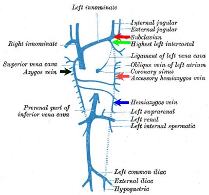
Gray's Anatomy
- The left superior intercostal vein may increase in size with the patient supine or during expiration
- The left superior intercostal vein may act as a collateral pathway, and therefore become distended, in patients with impending or actual superior vena caval obstruction, absence or obstruction of the inferior vena cava, congestive heart failure, congenital absence of the azygos vein (black arrow above) or increased portal venous pressure or, rarely, with partial or total anomalous pulmonary venous drainage
- It should not be mistaken for a left superior vena cava which, if present, may give rise to the superior intercostal vein rather than the left brachiocephalic vein
- If large enough, the aortic nipple could be mistaken for an aortic aneurysm or a mediastinal mass
- Imaging
- Conventional radiography
- Usually produces a rounded or pointed soft-tissue density of varying size adjacent to the lateral wall of the aortic knob on the frontal view
- It may project more superiorly or inferiorly along the border of the aortic knob or may sometimes be visible through the knob
- CT
- Contrast-enhanced chest CT images will show a curvilinear contrast-filled vessel along the left lateral border of the aorta that can usually be traced from the left brachiocephalic vein to the region of the accessory hemiazygos vein
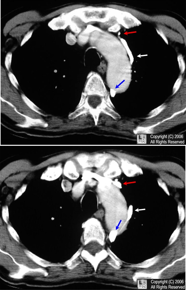
Left Superior Intercostal Vein. Contrast-enhanced chest CT images show a curvilinear contrast-filled
vessel along the left lateral border of the aorta (white arrow) that can be traced from the
left brachiocephalic vein (red arrow) to the region of the accessory hemiazygos vein (blue arrow)
For the same photo without the arrows, click here
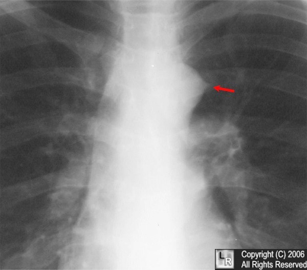 
Left. Close-up of aortic nipple on frontal chest radiograph (red arrow)
Right. Plate from Gray's anatomy showing venous drainage in chest.
McDonald C, Castellino R, Blank N: The aortic "nipple" the left superior intercostal vein. Radiology 96:533-536 1970
|
|
|
