|
|
Vanishing Lung Syndrome
Idiopathic Giant Bullous Emphysema
General Considerations
- Uncommon disorder
- Characterized by very large bullae
- Bullae are air-filled, thin-walled (<1mm) spaces in the lung resulting from destruction of alveolar tissue
- In vanishing lung syndrome the bulla takes up more than a third of the occupied lung
- Paraseptal emphysema and subpleural bullae are seen in virtually all patients
- Most also have separate centrilobular emphysema
- Most common in young men, mostly in smokers
Clinical Findings
Imaging Findings
- Bullous disease asymmetrically involved the upper lobes predominantly
- On high-resolution CT, bullae range from 1 to 20 cm in diameter, mostly in the range of 2-8 cm in diameter
- Bullous disease asymmetrically involved the upper lobes predominantly
Differential Diagnosis
- Mistaking a large bulla for a pneumothorax and inserting a chest tube into it will lead to a pneumothorax
- Find the visceral pleural white line to identify a pneumothorax and, when in doubt, do CT
Treatment
- Volume reduction surgery
- Extent of centrilobular emphysema is key variable for determining preoperative assessment of bullectomy
Complications
- Bullae compress adjacent lung and can predispose to pneumothorax


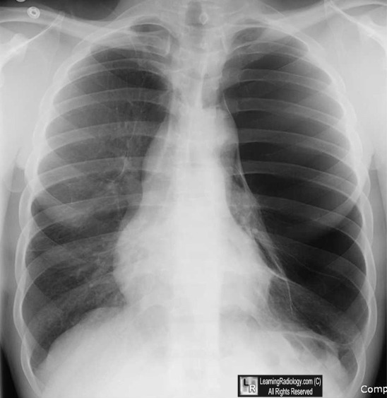
Vanishing Lung Syndrome. The chest radiograph shows a giant bulla occupying the
entire left hemithorax (white arrow).
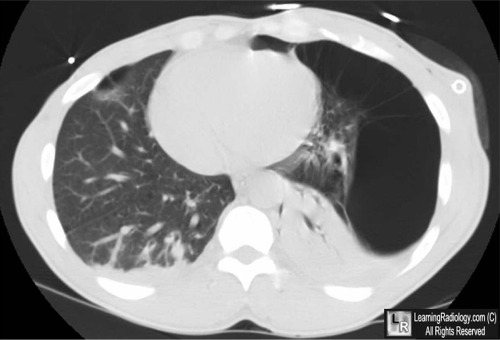
There is no visceral pleural line visible to indicate this is a pneumothorax.The CT scan of the lower chest shows the large bulla compressing
the lung medially and posteriorly (white arrows)
For these same photos without the arrows, click here and here
For more information, click on the link if you see this icon 
Idiopathic giant bullous emphysema (vanishing lung syndrome): imaging findings in nine patients. EJ Stern, WR Webb, A Weinacker and NL Muller. AJR, Vol 162, 279-282, 1994.
|
|
|
