|
|
Atlanto-Axial Subluxation/Dislocation
- Distance between the anterior surface of the dens and the posterior surface of the tubercle of C1 is usually 3 mm or less in adults and 5 mm or less in children
- This space is called by many names: predentate space, predental space, atlantodental distance
- The distance may increase slightly on flexion in children but is usually unchanged between flexion and extension in adults
- Forward movement of the atlas on the axis is normally restricted by the transverse ligament
- The transverse ligament is the primary restraint against atlantoaxial, anteroposterior movement
- Atlantoaxial instability is defined by an increase in the predentate space of greater then 3 mm in adults and 5 mm in children
- Symptoms will be present when the atlas moves far enough forward on the axis to narrow the spinal canal and impinge on the spinal cord
- The spinal canal is typically widest at the level of C2 and should not be less than 18 mm in ins widest AP dimension
- Non-traumatic conditions associated with increase in the atlantoaxial distance
- Down syndrome
- Due to laxity of the transverse ligament
- Grisel syndrome
- Atlantoaxial subluxation associated with inflammation of adjacent soft tissues of the neck
- Rheumatoid arthritis
- From laxity of the ligaments and destruction of the articular cartilage
- Osteogenesis imperfecta
- Neurofibromatosis
- Morquio syndrome
- Secondary to odontoid hypoplasia or aplasia
- Other arthridities
- While chronic atlantoaxial dislocations which occur in the above diseases may be severe yet asymptomatic, acute atlantoaxial dislocations are more often symptomatic and can be life-threatening
- Traumatic atlantoaxial subluxation/dislocation usually results from a motor vehicle collision in which an unrestrained occupant’s head strikes the windshield or dashboard
- The pathologic mechanism involves hyperflexion of the neck
- Almost all atlantoaxial dislocations involve forward movement of C1 on C2; posterior dislocation is extremely rare
- Anterior atlantoaxial dislocations may be, but are not necessarily, associated with a fracture of the dens (~50% at autopsy)
- Associated fractures of the skull and/or facial bones are common
- This injury is unstable
- Neurologic injury occurs from cord compression between the odontoid and posterior arch of C1
- Imaging findings
- Widening of the predentate space
- Disruption in the smooth curve of the imaginary line connecting the spinolaminar white lines of the vertebral bodies
- Soft tissue swelling
- C2 ─ retropharyngeal space should be < 7 mm
- C3 and C4 ─ retropharyngeal space should be < 5 mm, or
- Less than half the AP diameter of C3 or C4
- C6 ─ retrotracheal space should be < 22 mm in adults and < 14 mm in children (under15 years)
- Treatment
- These injuries are usually treated with some form of surgical fusion of C1 and C2
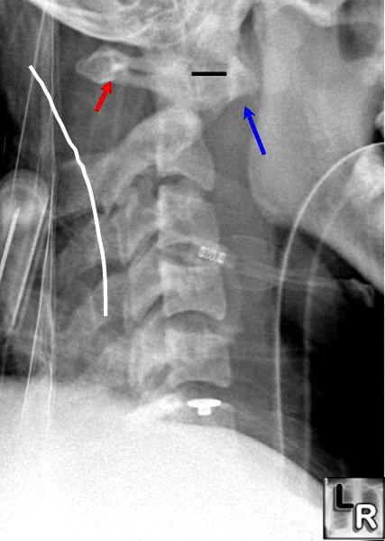
Atlantoaxial dislocation. Lateral view of the cervical spine done as a cross-table lateral
shows a marked increase in the distance between the anterior surface
of the dens and the posterior surface of the C1 tubercle (blue arrow) that measured 14 mm (black line),
well in excess of the 3 mm maximum in adults. The imaginary line connecting the spinolaminar
white lines (white line) shows that the body of C1 (red arrow) is displaced anteriorly relative to the remainder
of the spine. The patient died shortly after this study was obtained.
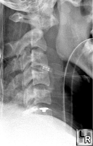
For a photo of the same image without the arrows, click here
Neck
Injuries:
II.
Atlantoaxial
Dislocation—A
Pathologic
Study
of
14
Traffic
Fatalities
Adams,
VI
Journal
of
Forensic
Sciences
37:2,
1992
|
|
|
