|
|
Dermatomyositis
Polymyositis
- Along with polymyositis, part of a group of idiopathic inflammatory myopathies with both cutaneous and visceral manifestations
- Affects the esophagus, lungs and heart
- Damaged chondroitin sulfate, atrophy of muscles, followed by calcification of muscle and subcutaneous tissue
- Most believe dermatomyositis is closely related to polymyositis although the pathogenesis of the two remain controversial
- Occur at age 5-10 and again in 50’s, dermatomyositis being the only one of the two diseases seen in children
- More common in females
- Linear and confluent calcifications in soft tissues of extremities
- Acro-osteolysis
- Chest-may have infiltrates associated, especially from aspiration
- Clinically
- Dysphagia
- Erythematous purple-red rash of eyelids, trunk and hands (seen in dermatomyositis)
- May be sole manifestation in up to 40% of patients with disease
- Painless, symmetric, proximal muscle weakness
- Associated with a higher incidence of malignancies of GI tract, lung, ovary , breast, kidney in adults, not usually children
- Imaging
- MRI may show an inflammatory myopathy
- Calcinosis universalis in dermatomyositis
- Diffuse cutaneous, subcutaneous and sometimes muscular calcification
- Usually affects children and young adults
- Not actual bone formation
- More linear than calcifications in scleroderma, which tend to be punctate (calcinosis circumscripta)
- Calcium-channel blockers have been reported to help in some cases of calcinosis
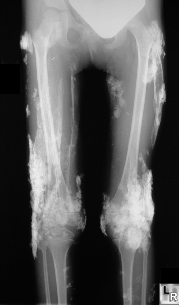
Dermatomyositis. Sheet-like calcifications seen in patients with
dermatomyositis is called calcinosis universalis because of its wide-spread distribution.
This is more likely to occur in younger patients with dermatomyositis.

Dermatomyositis. Multiple calcifications are seen in the subcutaneous tissue
on this cross-sectional CT scan of the abdomen in a patient with dermatomyositis.
- May resemble myositis ossificans progressiva
- Myositis ossificans progressive (fibrodyplasia ossificans progressiva)
- Begins with subcutaneous, painful masses in neck
- Progresses down back over shoulders, chest, abdomen
- Rounded or linear calcification starting in neck
- More clumplike in places than calcinosis universalis
- Ossification of voluntary muscles
- Treatment
- Prednisone
- Cytotoxic agents like methotrexate
- Prognosis depends on age, and cardiac, pulmonary or esophageal involvement
- Spontaneous remission has been reported in up to 1/5 of cases
|
|
|
