|
|
Posterior Strernoclavicular Dislocation
Important Points
- Sternoclavicular dislocations are rare, with the posterior subtype occurring less often than the anterior subtype.
- Complications occur in approximately 25% of posterior dislocations including pneumothorax, superior vena caval laceration, subclavian artery or vein occlusion, tracheal rupture or death.
- CT scans of the chest are most helpful in establishing the diagnosis although a serendipity view can provide a clue to its presence.
Discussion
- The sternoclavicular joint is a freely movable synovial joint.
- Normally, approximately 50% of the medial end of the clavicle articulates with the manubrium of the sternum.
- Sternoclavicular dislocations are one of the rarest of all dislocations to occur and require a considerable force involving a direct or indirect blow to the shoulder or shoulder region.
- The mechanism of injury most commonly occurs during a motor vehicle collision, followed in prevalence by athletic injuries or falls.
- In addition to sternoclavicular dislocations being a rare dislocation, the posterior subtype is even more rare.
- The anterior dislocation is nine times more common than a posterior dislocation.
- The mechanism of injury differs between the two subtypes, with the type of dislocation depending on the direction the shoulder is driven.
- If the shoulder is driven backwards, the clavicle moves forward as a fulcrum and an anterior dislocation results.
- If the shoulder is propelled forward, the clavicle moves posteriorly and a posterior dislocation results.
- Posterior dislocations can also result from a direct blow to the head of the clavicle.
Imaging
- The workup of a possible sternoclavicular dislocation is of utmost importance.
- A chest x-ray may be normal and additional views of the clavicle may be needed.
- The recommended view is a “serendipity view,” which is an AP view obtained by angling the x-ray tube 40 degrees cephalad with the patient in the supine position.
- Normally, the medial ends of the clavicles will appear at the same level, while a posteriorly dislocated clavicular head will appear lower, and an anteriorly dislocated clavicular head will appear higher than its counterpart on the serendipity view.
- CT is the preferred imaging modality because of its ability to easily demonstrate the dislocation as well as soft issue abnormalities
Complications
- Complications are rare with anterior dislocations but occur in approximately 25% of posterior dislocations.
- These complications include pneumothorax, superior vena caval laceration, subclavian artery or vein occlusion, tracheal rupture or death.
Treatment
- Posterior dislocations produce a unique management problem.
- While acute dislocations may be treated non-operatively, closed reductions of posterior dislocations may fail or produce additional complications due to mediastinal injury.
- Therefore treatment options include open reduction and internal fixation if additional stabilization is required.
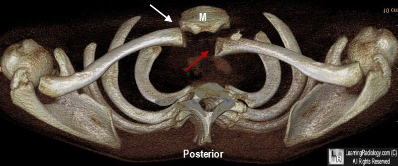
Posterior Dislocation of the Left Sternoclavicular Joint. A 3D reconstruction in the axial plane shows posterior dislocation of the medial head of the left clavicle Red arrow) relative to the manubrium of the sternum (M). The opposite SC joint is intact (white arrow).
Rudzinski J, Burelbach J. Sternoclavicular Joint Injury, eMedicine, March 10, 1995.
Brinker MR, Simon RG. Pseudo-dislocation of the sternoclavicular joint. J Orthop Trauma 1999 Mar-Apr; 13(3): 222-5.
Cope R: Dislocations of the sternoclavicular joint. Skeletal Radiol 1993; 22(4): 233-8.
Thomas DP, Davies A, Hoddinott HC: Posterior sternoclavicular dislocations--a diagnosis easily missed. Ann R Coll Surg Engl 1999 May; 81(3): 201-4.
|
|
|
