|
|
Osteoma of the Paranasal Sinus
General Considerations
- Most common tumor of the paranasal sinuses
- Most frequently seen in the frontal and ethmoid sinuses
- Benign tumor of membranous bone consisting of dense, compact bone
- Majority of paranasal osteomas are discovered serendipitously
- In the skull, they usually arise from the outer table
Clinical Findings
- Most are asymptomatic
- Rarely, large osteoma in the frontal or ethmoid region may displace globe forward and cause proptosis
- Obstruction of a sinus ostium may lead to infection or formation of a mucocele
- Very rarely, an osteoma may erode through the dura leading to cerebrospinal fluid rhinorrhea or intracranial infection
Imaging Findings
- Well-circumscribed, sharply-marginated round and very dense lesions usually less than 2 cm in size
- Usually grow into the sinus
- Multiple paranasal osteomas are found in Gardner’s syndrome
- Multiple osteoma of the mandible and maxilla, along with the frontal, sphenoid and ethmoid sinuses, rarely the long bones or phalanges
- Cutaneous and soft tissue tumors
- Association between colonic polyps with a predilection to malignant degeneration
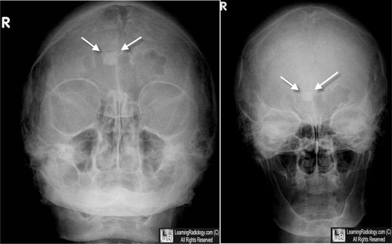
Osteoma of the Frontal Sinus. Two frontal views of the skull demonstrate an incidental rounded, sclerotic lesion growing into the right frontal sinus (white arrows).
|
|
|
