|
|
Pericardial Cyst
- Fluid-filled cysts of the parietal pericardium consisting of a single layer of mesothelial cells
- Usually discover at age 30-40 years, predominantly in males (3:2)
- Most are asymptomatic and incidental findings
- Atypical chest pain can occur
- They are usually (75%) located at the cardiophrenic angle almost always on the right (3:1)
- DDX of a right cardiophrenic angle mass
- Pericardial cyst
- Sequestration
- Foramen of Morgagni hernia
- They can occur higher and may extend into major fissure
- Classically they are soft and can be flattened on the edge that faces the fissure
- They rarely occur in the mediastinum
- Imaging findings
- Sharply marginated
- Round or oval mass
- From 3-8 cm in size usually
- They can change in size and shape with respiration or body position
- Rarely calcify
- On CT, their attenuation values of 20-40 HU, occasionally higher
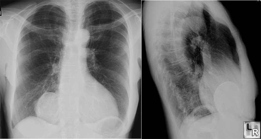
Pericardial Cyst. Frontal and lateral views of the chest demonstrate a mass at the right cardiophrenic angle with rim-like calcification that indicates the calcification has formed in the wall of a hollow viscus. This is a characteristic location for a pericardial cyst, which is calcified in this case.
|
|
|
