|
|
Posterior Hip Dislocation
General Considerations
- Hip dislocation accounts for only 5% of all dislocations
- Posterior hip dislocations are much more common than anterior hip dislocations (90% to about 10%)
- Mechanism in posterior dislocation classically is unrestrained occupant of a motor vehicle accident, especially collisions which are head-on, in which the flexed knee strikes dash with hip flexed and adducted
- Force is transmitted from the foot or ankle along femoral shaft to the hip
- Associated with fractures of the posterior rim of the acetabulum
- Posterior dislocations can also result from falls from a height
- Anterior dislocations are more apt to occur if the hip is abducted at the time of injury
- Greater force required to dislocate an adult's hip than a child's
Clinical Findings
- Affected limb is shortened, adducted, and internally rotated, with the hip and knee in slight flexion
- Signs of vascular or sciatic nerve injury
- Pain in hip, buttock, and posterior leg
- Loss of sensation in posterior leg and foot
- Loss of dorsiflexion (peroneal branch) or plantar flexion (tibial branch)
- Loss of deep tendon reflexes (DTRs) at the ankle
- Local hematoma
Imaging findings
- Conventional radiography
- In posterior dislocations, the head of the femur is usually situated superior and lateral to its normal position in the acetabulum
- In anterior dislocations, the head usually rests inferior and medial to its normal acetabular position
- May be subtle if head in AP plane appears as if it still resides in the acetabulum
- There may be associated fractures of the head of the femur and/or posterior rim of the acetabulum
- The posterior rim of the acetabulum normally is the more lateral of the two edges (anterior rim and posterior rim) seen on the anteroposterior (AP) view of the pelvis
- Since the posteriorly dislocated head lies closer to the cassette, the posteriorly dislocated head may appear smaller than the head on the opposite side which lies farther from the cassette and is magnified more
- Computed tomography (CT)
- Provides an accurate means of evaluating not only the dislocation but the associated fractures as well
Treatment
- Requires immediate pain management
- Reduction of the dislocation within 6-12 hours
- Avascular necrosis of the femoral head is more likely to occur if the reduction does not occur before 6 hours
Prognosis
- Good-to-excellent results in 76-93% of patients

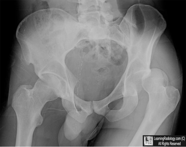
Posterior Hip Dislocation. The left femoral head (white arrow) lies superior (and posterior) to the acetabulum (blue arrow). No Pelvic fracture is seen.
For this same photo without the arrows, click here
For more information, click on the link if you see this icon 
Posterior Hip Dislocations. EmEdicine. J Naradzay, MD and P Carter, MD.
|
|
|
