|
Ascites
· Causes
o Cirrhosis
o Hypoproteinemia
o Chronic renal failure
o Carcinomatosis
o Polyserositis
o Pancreatitis
o TB peritonitis
o Meig’s syndrome
o Constrictive pericarditis
o Budd-Chiari syndrome
· Imaging Findings
o Conventional radiographs
§ Uniform grayness to abdomen
§ Central placement of bowel loops
§ Separation of adjacent loops
§ Loss of definition of the liver and/or spleen edge
§ Bladder-ears ─ fluid collects in pelvis on either side of bladder in peritoneal space
§ Thickening of peritoneal flank stripe
§ Medial displacement of ascending and descending colon
§ Bulging flanks
o On CT
§ Sparing of the “bare” area of the posterior aspect of the right lobe of the liver which is not covered by peritoneum
· Fluid that lies posterior to the liver at this point is pleural effusion, not ascites
§ Ascitic fluid lies anterior to the diaphragm on axial sections, pleural fluid is posterior
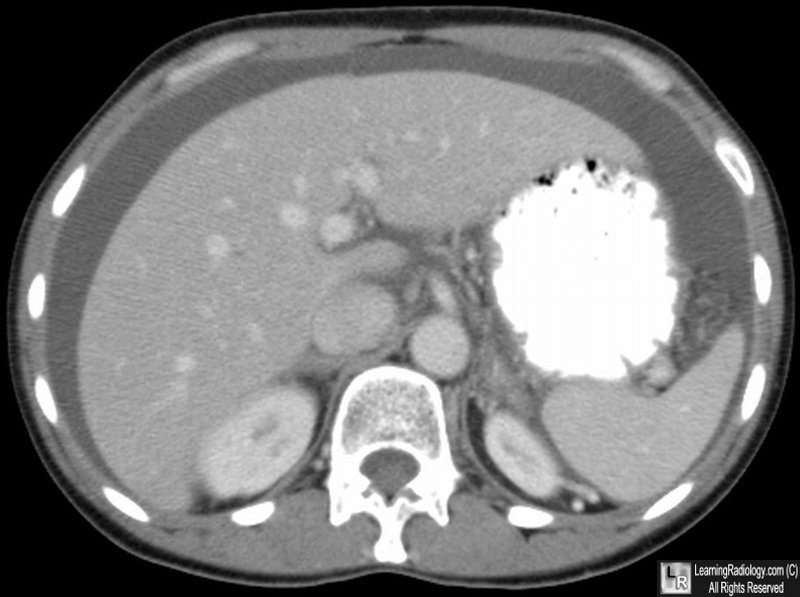
Ascites. Axial CT scan of the abdomen shows low density ascitic fluid surrounding the liver, spleen and stomach.
| 