|
|
Jefferson Fracture
General Considerations
- Burst fracture of the ring of C1
- Typically caused by an axial-loading force on the occiput of the head
- Classically, it involves fractures of the anterior arch of C1 on both the right and left sides and the posterior arch of C1 on both the right and left sides (4 fractures)
- But fracture variants may include two or three-part fractures
- There is usually no associated neurologic deficit as the ring of C1 widens when it fractures limiting cord compression
Mechanism
- Original description in 1920 by Sir Geoffrey Jefferson, an English neurologist and neurosurgeon, in “Fracture of the atlas vertebra: report of four cases, and a review of those previously recorded that appeared in the British Journal of Surgery
- He described the role of axial compression
- Today, this most frequently occurs when diving into shallow water, the head strikes an obstacle (or the bottom of the pool) and the force is transmitted to the cervical spine
- It may also occur from motor vehicle accidents in which the head is thrown forcefully against the windshield, frequently producing both hyperextension and compression
- Another mechanism is falling onto the head from a height
Associated injuries
- Approx 1/3 of Jefferson fractures are associated with a fracture of C2
Clinical findings
- Patients usually complain of upper neck pain following trauma
- Neurological examination is usually normal
Imaging findings
- Conventional radiography
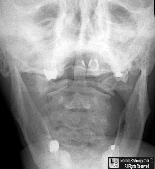
- Open-mouth (odontoid) view is the most revealing
- Classically there is bilateral, lateral offset of C1 on C2
- Lateral view:
- May show prevertebral soft tissue swelling anterior to C1
- Pre-dentate space (distance between the anterior tubercle of C1 and the dens) may be widened to greater than 3 mm if there is damage to the transverse ligament
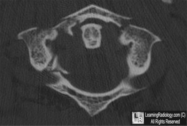
- CT Scan
- Demonstrates the number of fractures, their locations and degree of displacement of fragments
Treatment
- Depends in part on whether there is damage to the transverse ligament and the degree of offset of C1 on C2
- Treatments include collar or brace for 3 months all the way through cranial traction


Jefferson Fracture. An open mouth view of the cervical spine (upper) shows bilateral lateral
offsets of the lateral masses of CI on C2 (white arrows). This is presumptive evidence of a Jefferson fracture. The axial CT scan (lower) confirms the burst fracture of the arch of C1 bilaterally in the anterior arch and on the right in the posterior arch (white arrows)
For these same photos without the arrows, click here and here
For more information, click on the link if you see this icon 
Wheeless’ textbook of Orthopaedics
Mark R Foster, MD, PhD eMedicine
|
|
|
