|
|
Avascular Necrosis of the Lunate
Kienbock Disease, Lunatomalacia
Submitted by Seema Hasan, MD
- Avascular necrosis of lunate bone
- Predisposed
- Individuals engaged in manual labor with repeated or single episode of trauma
- Frequency
- Usually affects men aged 20-40 yrs.
- Mostly unilateral
- More often in right wrist
- Pathophysiology
- Vascular impairment due to acute or chronic injury
- Lunate develops osteonecrosis due to loss of blood supply, causing pain and stiffness in the wrist
- In late stages, the bone collapses eventually leading to degenerative changes and osteoarthritis in the radiocarpal joint
- Clinical findings
- Progressive pain
- Soft-tissue swelling of wrist
- Imaging findings
- The disease can be staged based on radiographic findings
- Lichtman's Radiographic Classification of Kienbock's Disease
- Stage I - Normal radiograph
- Stage II - Sclerosis of lunate with possible decrease of lunate height on radial side only
- Stage IIIa - Lunate collapse, no scaphoid rotation
- Stage IIIb - Lunate collapse, fixed scaphoid rotation
- Stage IV - Degenerative changes around the lunate
- The disease may also be associated with negative ulnar variance
- Bone scan and MRI may be helpful early in the course of the disease when there are minimal radiographic findings
- Treatment
- Initial therapy is conservative management
- Anti-inflammatory medications and splinting or casting
- Operative treatment is based on the stage of the disease and may involve
- Revascularization procedures
- Ulnar lengthening or radial shortening
- Fusion or excision of carpal bones
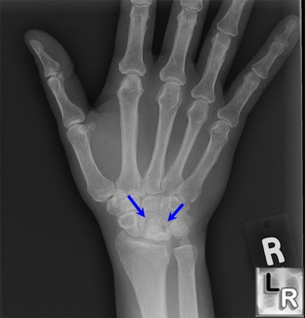
Kienbock Disease. Frontal view of the
hand and wrist
demonstrates
sclerosis,
irregularity and collapse of the lunate (blue arrows).
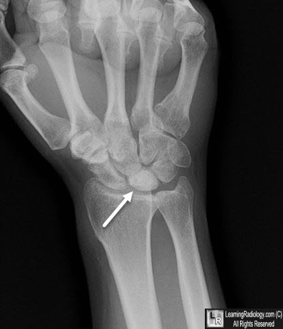
Kienbock Disease. Frontal view of the
hand and wrist
demonstrates
sclerosis of the lunate (white arrow).
emedicine,"Kienböck Disease" : Article by Brian J Divelbiss, MD
Wheelessonline.com, "Kienbock's disease: Lunatomalacia"
|
|
|
