|
|
Ulcerative Colitis
Pathology
- Predominantly mucosal disease, possible auto-immune producing crypt abscesses
- Usual age at onset is 20-40; another peak at 60-70
- Equal male:female ratio
Clinical
- Recurrent episodes of bloody diarrhea
- Electrolyte depletion
- Abdominal pain
- Fever
- Periods of exacerbation and remission
- Iritis, erythema nodosum, pyoderma gangrenosum
- Pericholangitis, chronic active hepatitis, sclerosing cholangitis, fatty liver
- Spondylitis, peripheral arthritis, RA (10-20%)
- Thrombotic complications
Location
- Begins in rectum with retrograde progression
- Rectosigmoid involved 95%; continuous involvement of left colon
- Terminal ileum in 5-10% with backwash ileitis
Imaging Manifestations
- Acute inflammatory stage
- Spasm and irritability
- Fine mucosal granularity=earliest finding on air-contrast BE
- Spiculated, serrated bowel margins from tiny, multiple ulcerations
- Collar button ulcers-from undermining (not specific for UC)
- Double-tracking=long, longitudinal ulcers in submucosa
- Thumbprinting=from edema of wall
- Pseudopolyps=scattered islands of edematous mucosa in a sea of ulcerated mucosa
- Widening of the presacral space
- Subacute stage
- Coarser, more granular mucosa
- Inflammatory polyps= frondlike lesions of inflamed mucosa
- Chronic stage
- Shortening of the colon=may be from spasm of longitudinal muscles or from irreversible fibrosis (lead-pipe colon)
- Loss of haustrations on left side of colon
- Postinflammatory polyps=filiform polyps=long worm-like lesions
- Backwash ileitis (5-10%)=wide open ileocecal valve and dilated terminal ileum
Differential Diagnosis
- Crohn’s disease–skip lesions: R colon; TI abnormal
- Cathartic colon-loss of haustrations on Right side of colon; rectum spared
- Familial polyposis–multiple polyps but no inflammatory changes
- Radiation ileitis–should have other loops involved and appropriate hx
- Lymphoma–should have tumor masses, less spasm
- Amebiasis–cone-shaped cecum
Extra-intestinal Manifestations
- Fatty infiltration of the liver
- Gallstones (28-34%)
- Sclerosing cholangitis
- Bile duct carcinoma
- Amyloidosis
- Urolithiasis:oxalate/uric acid stones
- Migratory arthritis
- Sacroiliitis and ankylosing spondylitis
- Erythema nodosum and uveitis
Complications
- Toxic megacolon
- Adenocarcinoma of the colon (1-16%)
- Increased risk of developing ca of colon with long-standing (usually more than 25 years) ulcerative colitis
- Higher incidence of multiple carcinomas
- Usually involve distal transverse colon, descending colon and rectum
- May present with smooth, tapering edges and resemble a benign stricture or may be annular constricting lesions
- Colonic strictures (10%)
- Smoothly tapering edges, usually single, commonly in sigmoid; must be differentiated from ca
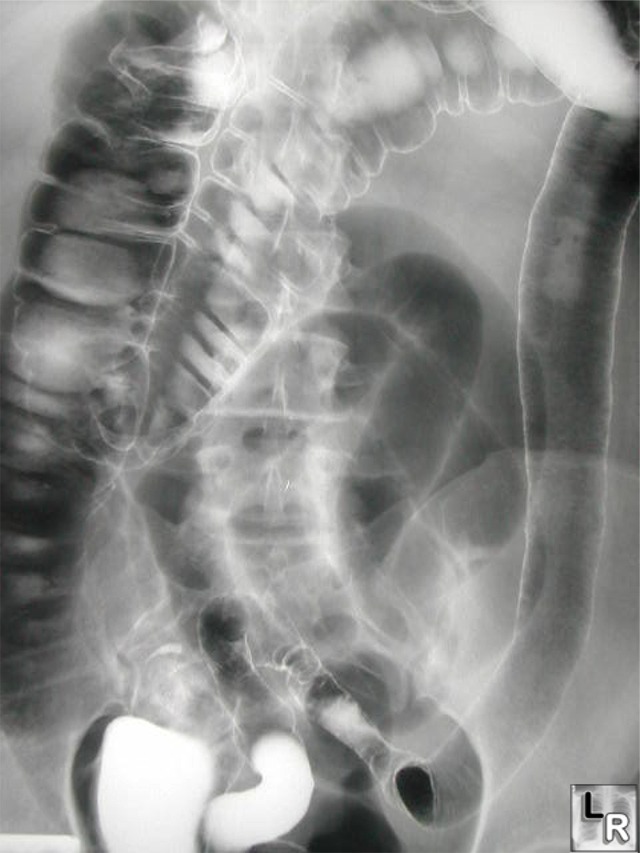
Ulcerative Colitis: Barium enema
examination
demonstrates loss of
haustral folds in the
entire descending
colon with small
ulcerations suggested.
The colon has a
"lead-pipe"
appearance.
The distribution and
appearance are
suggestive of
ulcerative colitis.
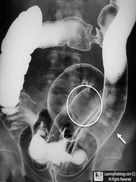
Ulcerative Colitis: Barium enema
examination
demonstrates loss of
haustral folds in the
entire descending
colon (white arrow) with a granular appearance to the mucosa suggesting small
ulcerations.
The distribution and
appearance are
suggestive of
ulcerative colitis.
|
|
|
