|
|
Renal Papillary Necrosis
General Considerations
- Necrosis of the renal medullary pyramids and papillae with many causes, all of which mediate the development of ischemia
- Infection is frequent finding, contributing to the clinical presentation of with fever and chills in about 2/3 of patients and positive urine cultures in 70%
- But papillary necrosis can also develop without infection being present
- Inflammatory reaction in the interstitium of the kidney compresses and compromises the medullary vasculature and predisposes the patient to ischemia and papillary necrosis
- Other diseases can also impair this circulation, among them
- Diabetes mellitus
- Urinary obstruction
- Analgesic nephropathy
- Phenacetin, with its toxic metabolite, p-phenetidin
- Also occurs with NSAIDS (non-steroidal anti-inflammatory drugs)
- But usually with another predisposing factor present
- Any condition associated with ischemia predisposes a person to papillary necrosis, such as
- Shock
- Dehydration
- Hypovolemia
- Sickle cell disease
- Tuberculosis
- Trauma
- Cirrhosis = alcoholism
- Coagulopathy
- Renal vein thrombosis
- Hemophilia
- Christmas disease
- Acute tubular necrosis
- Most patients who develop papillary necrosis have two or more contributing factors
- Usually bilateral
- Can affect a single papilla or entire kidney may be involved
- Mean age of onset is 53 years
- More than 90% of cases occur in individuals older than 40
- Uncommon in patients younger than 40 and in the pediatric population
- More often in women than in men
- Types
- Clinical findings
- Fever and chills
- Flank and/or abdominal pain
- Hematuria
- Acute ureteral obstruction from sloughed papillae manifests as flank pain and colic from hydronephrosis or pyonephrosis
- Hematuria is almost always present
- Clinical picture in such cases may also include fever, chills and sepsis.
- Imaging findings
- The kidneys are usually normal in size until they contract in the late stages of the disease
- Linear streak of contrast may appear inside of calyces representing void left by sloughed papilla (lobster claw sign)
- Widening of the fornices from shrinking of the papillae
- Larger collection of contrast may fill cavities inside of calyces representing a calyx without a papilla
- Ring shadows can develop in the medulla outlining detached papilla within contrast material-filled cavity
- Often in a triangular shape, referred to as the ring sign
- Sloughed papillae can produce filling defects in internal collecting system or ureters
- The ring shadow or sloughed papilla can rarely calcify
- Complications

Renal Papillary Necrosis. Image from a CT Urogram shows numerous irregular collections of contrast arising
from the calyces, some streak-like densities and overall distortion of the normal medullary-calyceal anatomy (yellow circles).
For this same photo with the arrows, click here
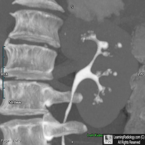
Renal Papillary Necrosis. Image of left kidney from a CT Urogram shows numerous irregular collections of contrast arising
from the calyces, some streak-like densities and overall distortion of the normal medullary-calyceal anatomy.
For more information, click on the link if you see this icon 
eMedicine. JM Donohoe, JH Mydlo, AN Khan, M Chandramohan, and S Macdonald
|
|
|
