|
|
Pseudotumor of Lung
Vanishing Tumor
General Considerations
- Sharply marginated collection of pleural fluid contained either within an interlobar pulmonary fissure or in a subpleural location adjacent to a fissure
- Result from transudation from the pulmonary vascular space
- Commonly manifest as incidental radiographic findings in patients with congestive heart failure
- Other causes of transudates include
- Hypoalbuminemia
- Renal insufficiency
- Patients who develop pseudotumors tend to develop them repeatedly when the underlying condition causing the transudate (like CHF) returns
- Pseudotumors may be erroneously diagnosed as parenchymal lung nodules or masses
- Presence of an interlobar pleural effusion does not always correspond to the severity of the left heart failure
Imaging findings
- Lenticular or biconvex contour
- Located along the course of interlobar fissures
- Most occur in the minor (horizontal) fissure (more than 75%) and are seen on both the frontal and lateral radiograph
- Those that occur in the oblique or major fissure may only be seen on the lateral view well
- Infrequently, they occur in the horizontal and oblique fissures simultaneously
- Most are less than 4 cm in size
Management
- The underlying condition is managed
- Typically an incidental finding that has minimal impact on patient management
- Occasionally, it may be the only sign of cardiac decompensation
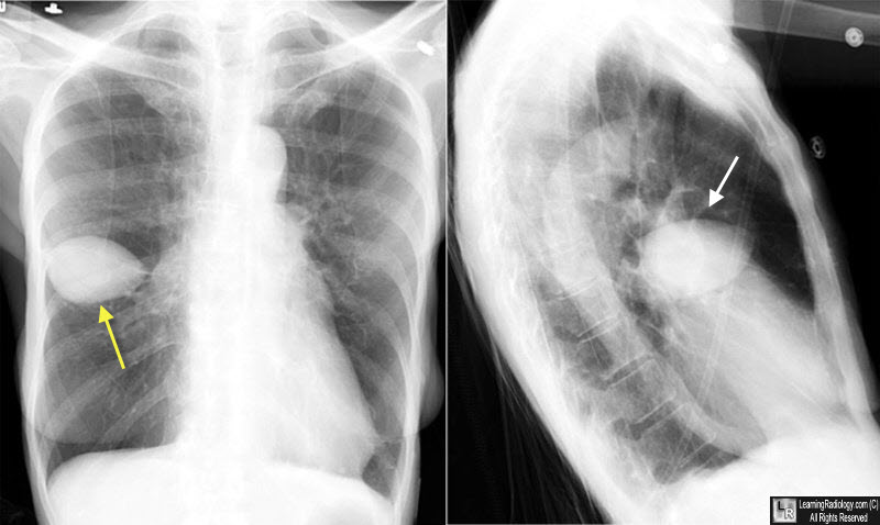
Pseudotumor of Lung. Frontal and lateral views of the chest demonstrate a lemon-shaped soft-tissue density corresponding to the location of the minor fissure on both views (white arrows). This is a classic appearance for a pseudotumor of the lung.
Massive Pulmonary Pseudotumor: Brian M. Haus, BA; Paul Stark, MD; Scott L. Shofer, MD and Ware G. Kuschner, MD, FCCP Chest. 2003;124:758-760
|
|
|
