|
|
Thyroid Ophthalmopathy
- Most common cause of
proptosis in adults
- Typically occurs from
20-50 years old
- Patients are usually
hyperthyroid, but may be euthyroid
- Neuroimaging usually reveals thick
muscles with tendon sparing
- Inferior rectus and
medial rectus muscles are most commonly
involved
- Usually bilateral, but
may be asymmetric
- Grave’s Disease =
Diffuse Toxic Goiter
- Autoimmune disorder
with thyroid stimulating antibodies (LATS)
producing hyperplasia and hypertrophy of
thyroid gland
- Age of incidence
- 3rd-4th decade
- Female predominance
7:1
- Laboratory findings
- Elevated T3 and T4
- Depressed TSH
production
- Clinical findings
- Pretibial myxedema
- Ophthalmopathy
- Periorbital edema
- Lid retraction
- Opthalmoplegia
- Proptosis
- Malignant
exophthalmos
- Diffuse thyroid
enlargement
- US
- Identical to diffuse
goiter
- Global enlargement of
2-3 times normal size
- Normal and/or
diffusely hypoechoic pattern
- Hyperemia on color
Doppler
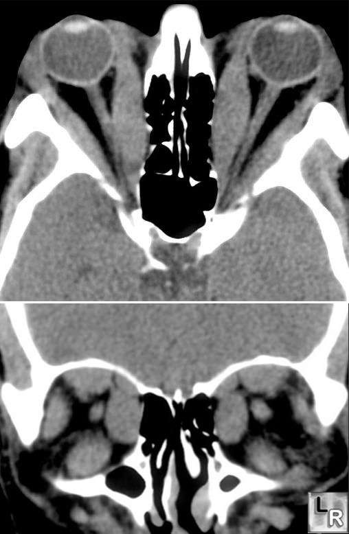
Thyroid Ophthalmopathy. Axial and coronal CT scans of the orbits show
marked enlargement of the
extraocular muscles with sparing of the tendons
consistent
with the ophthalmopathy seen with Grave's disease
|
|
|
