|
|
Impacted Chicken Bone in Esophagus
- Soft tissue
measurements on the lateral neck image
- At C3: <3 mm (less than
1/3 AP diameter)
- At C6: < the AP width of
C6 vertebral body
- Impacted esophageal foreign bodies
- Food or true foreign
bodies
- Chicken bones
(opaque), fish bones (non-opaque)
- Coins, toy trucks
- Most often they impact
just below cricopharyngeous (70%)
- Another 20% impact at
the level of the aortic arch
- Another 10% at EG
junction
- Once past the
esophagus, most foreign bodies will pass
through the GI tract
- Clinical findings of an
impacted esophageal foreign body
- Dysphagia and
odynophagia most commonly
- Even if FB passes,
many complain of pain referable to cervical
esophagus
- Imaging Findings
- Always check for lead
lines in children
- Chicken bones are
usually opaque
- Fish bones contain
less calcium and usually are not
- Plain films usually do
not demonstrate the FB but are still obtained
first
- If negative, then
either contrast esophagram or CT if high
index of suspicion
- Treatment
- Removal is most often
performed using endoscopy
- Temporization and
surgery are other options
- An ingested button
battery lodged in esophagus must be removed
immediately
- Complications of an
impacted foreign body
- Perforation
- Longer the FB remains
impacted (>24hrs), higher incidence of
perforation
- Stricture
- Diverticulum formation
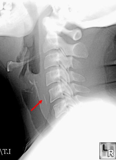
Impacted Foreign Body. Lateral radiograph of the neck
demonstrates a linear density in the region of the
proximal esophagus (red arrow) consistent with an
impacted foreign body--in this case, a chicken bone.
There is no air in the soft tissues and no soft
tissue swelling is
identified to indicate the presence of a
retropharyngeal abscess.
|
|
|
