|
|
Ranula
Submitted by Tony Chang, MD
- Mucus retention cyst that occurs in the sublingual gland
- Ranulas do not communicate with the duct
- Etiologies include
- Prior trauma, usually iatrogenic from prior surgery
- Obstruction of the sublingual gland or its duct
- Two types of ranulas
- Simple ranulas are true cysts
- Occur in floor of the mouth above the level of the mylohyoid with a lining formed by the sublingual gland capsule
- Plunging/deep/diving ranulas
- Occur when the simple ranula ruptures and is walled off by an inflammatory response
- Plunging ranulas are pseudocysts partially contained by the remaining epithelium and inflammatory cells that react to irritative saliva
- Plunging ranulas usually extend below the level of the mylohyoid
- Clinical findings
- Both types present as a painless mass in the sublingual space
- Plunging ranulas extending inferiorly into the submental or submandibular space
- Imaging findings
- Typical CT findings include
- Smooth, well delineated, cystic lesion
- Splaying the genioglossus and mylohyoid with a uniformly thin, non-enhancing wall
- Ranulas can be slightly increased in attenuation especially the higher the protein content within the fluid
- Infected ranulas may have thickened enhancing walls and cannot be distinguished from an abscess
- MRI findings
- Low signal intensity on T1 weighted imaging
- High signal intensity on T2 weighted imaging but varies with protein content
- Treatment
- Simple ranulas usually treated with transoral drainage and excision of ipsilateral sublingual gland
- Plunging ranulas may require more extensive surgical neck dissection
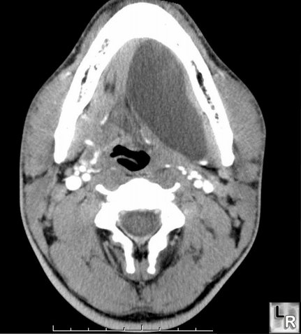
Ranula. CT scan shows a large mucous
retention cyst arising from the sublingual gland (ranula)
Som and Curtin, Head and Neck Imaging
|
|
|
