|
|
Choroid Plexus Cysts
Submitted by Jonathon Dorff, MD
- Cyst-like spaces that occur in the
choroid in approximately 1-6% of fetuses between 13 and
24 weeks gestation
- Majority are small and incidental,
disappearing by 26 weeks gestation
- Thought to represent entrapment of
cerebrospinal fluid within an in-folding of neuroepithelium
- May be associated with chromosomal
abnormalities, especially trisomy 18
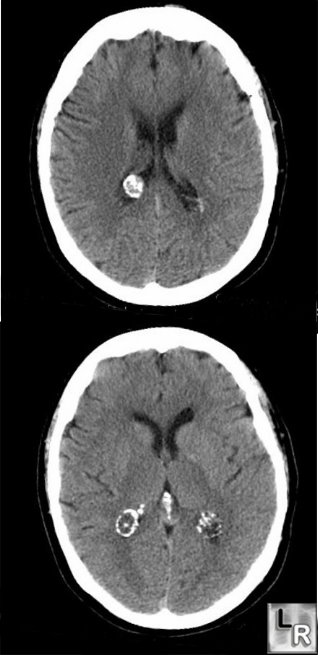
Choroid Plexus Cysts. Two images form an unenhanced axial CT
of the brain show ring-like calcifications in the region of the choroid plexus representing
choroid plexus cysts
Middleton, William D., etc: Ultrasound: The Requisites, 2nd edition, 2004.
Rumack, Carol, etc: Diagnostic Ultrasound, 3rd edition,
2005.
|
|
|
