|
|
Chilaiditi’s Syndrome
Hepatodiaphragmatic Interposition of the Intestine
General Considerations
- Pronounced “Ky-La-Ditty”
- Refers to the usually asymptomatic interposition of the bowel (usually hepatic flexure of the colon) between the liver and the (right) hemidiaphragm
- Seen in 0.1-0.25% of chest x-rays
- Most frequently an incidental finding
- More often in males
- Almost always in adults
- May be present intermittently
- Factors contributing to its occurrence include
- Absence of normal suspensory ligaments of the transverse colon
- Abnormality or absence of the falciform ligament
- Redundant colon, as might be seen with chronic constipation or in bedridden individuals
- Aerophagia
- Paralysis or eventration of the right hemidiaphragm
- Patients with chronic lung disease, cirrhosis and ascites
- The “sign” refers to the usually asymptomatic presence of the interposed bowel
Clinical Findings
- The “syndrome” may involve
- Abdominal pain
- Constipation
- Vomiting
- Respiratory distress
- Anorexia
Imaging Findings
- Chilaiditi’s Syndrome is important because it can simulate pneumoperitoneum
- Look for the presence of haustral folds which can establish the air beneath the diaphragm is contained within large bowel
- Left lateral decubitus abdominal films may help in this distinction
- Concomitant pneumoperitoneum may be more difficult to diagnose


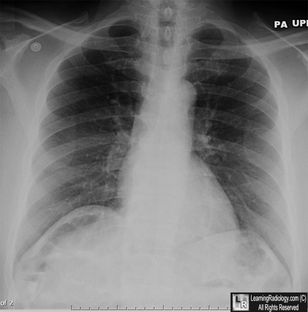
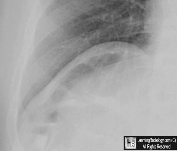
Chilaiditi's Syndrome. Top: There is interposition of colon between the right
hemidiaphragm and the liver producing a crescentic lucency in the right upper quadrant
(white arrow) that can be mistaken for free air. Bottom: Look for the haustral folds in the lucency
(yellow arrows) to help establish the air is in the colon.
For these same photos without the arrows, click here and here
For more information, click on the link if you see this icon 
|
|
|
