|
|
Metastatic Disease to Bone
Osteoblastic, Osteolytic
- Metastases are most common malignant
bone tumors
- Most involve axial skeleton
- Skull, spine and pelvis
- Rarely do mets occur distal to elbows
or knees
- Spread hematogenously
- Most frequently occur where red bone
marrow is found
- Mets to spine frequently destroy
posterior vertebral body including pedicle first=”pedicle-sign”
- 90% of skeletal mets are multiple
- Primary carcinomas that frequently
metastasize to bone
- The next four lesions comprise 80% of
all metastases to bone
- Breast (70% of bone mets in women)
- Lung
- Prostate (60% of all bone mets in
men)
- Kidney
- Also
- Thyroid
- Stomach and intestines
- Clinical
- Most lesions are asymptomatic
- When symptomatic, pain is major
symptom
- Fractures of the lesser trochanter in
adults should be considered pathologic until proven
otherwise
- Imaging Findings
- In general, mets have little or no
soft tissue mass associated with them
- Usually no periosteal reaction
- May appear as moth-eaten, permeative
or geographic lesions
- Indistinct zones of transition
- No sclerotic margins
- May be expansile
- Soap-bubbly (septated)
- May be sharply circumscribed or have
indistinct borders
- Metastases that are typically purely
lytic
- Metastases that are usually mixed
lytic and sclerotic
- Metastases that are usually purely
blastic
- Prostate
- Medulloblastoma
- Bronchial carcinoid
- No matter what the primary, skull
metastases are usually lytic in appearance
Most Common Tumors to Metastasize to Bone (80% of bone mets) |
| Tumor |
Appearance |
Prostate |
Blastic |
Breast |
Mixed |
Lung |
Predominantly lytic |
Renal Cell Ca |
Predominantly Lytic |
- Imaging findings suggestive of a
particular primary tumor
- Lesions distal to elbows and knees
- 50% are from lung and breast
- Expansile and lytic (soap-bubbly)
- Diffuse skeletal sclerosis or
multiple round, well-circumscribed sclerotic lesions

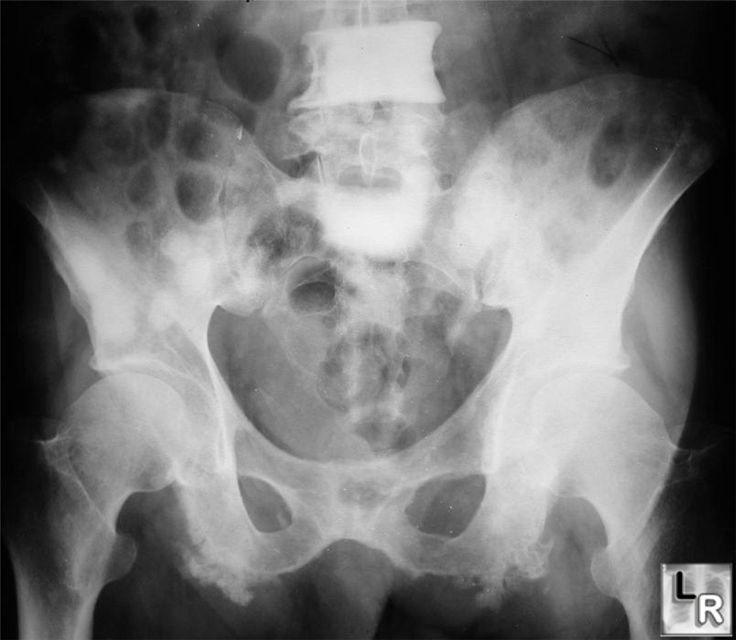
Multiple osteoblastic metastases to the
pelvis and lumbar vertebral bodies from carcinoma of the
prostate
Note discrete rounded sclerotic lesions in right ilium and
"ivory vertebra" involving
L4 and S1.
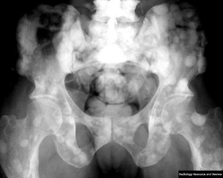
Multiple osteoblastic metastases to the
pelvis and lumbar vertebral bodies from carcinoma of the
prostate. Another case again shows innumerable, rounded sclerotic lesions throughout pelvis and femurs and an
"ivory vertebra" involving
L4 and S1.
- Cookie-bite lesions of the cortices of long
bones
- Radioscintographic studies
- Bone scans are extremely sensitive
but not very specific
- 10-40% of lesions will not be
visible on plain film but will be positive on bone scans
- CT or MRI can be used to show
findings in patients with negative conventional
radiographs and positive bone scans
- Complications of metastases to bone
- Pathologic fractures
- Destruction of 50% or more of bone
suggests impending pathologic fracture
- Spinal cord compression
- Treated lytic mets may become
sclerotic with treatment
Orthopedic Radiology: A Practical Approach, Greenspan, Adam; Lippincott, 2000
Diagnosis of Bone and Joint
Disorders, Resnick, Donald, W. B. Saunders
Musculoskeletal Imaging: The Requisites, Manaster, BJ et al; Mosby, 2002
|
|
|