|
|
Esophageal Web
- Ringlike constriction of upper esophagus covered on superior and inferior surfaces by squamous epithelium
- Three types have been described:
- A non-specific or idiopathic web (most common)
- Webs associated with Plummer-Vinson Syndrome
- Webs associated with epidermolysis bullosa dystrophica or graft-versus-host disease
- Usually found in middle-aged females
- Plummer-Vinson Syndrome=Patterson-Kelly syndrome
- Iron deficiency anemia
- Stomatitis
- Glossitis
- Dysphagia
- Spoon-shaped nails
- Esophageal webs
- Some question as to whether such a syndrome exists
- Location
- Cervical esophagus anteriorly at level of the cricopharyngeous (C5-C6)
- Best visualized with maximal distension
- Distal esophageal webs may arise from gastroesophageal reflux
- Imaging Findings
- Thin, transverse filling defects
- Perpendicular to anterior esophageal wall
- Usually less than 3mm in thickness
- Frequently they are not circumferential
- Increased risk of upper esophageal carcinoma
- DDx
- Prominent cricopharyngeous muscle
- Arises posteriorly at C5-C6 and produces a much broader defect
- Stricture
- Treatment
- Balloon dilatation
- Bougienage during esophagoscopy
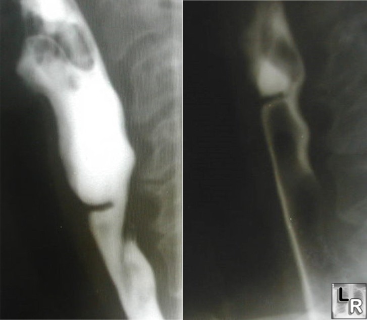
Esophageal Web. Barium esophagram demonstrates a thin membrane
arising from the anterior wall of the cervical
esophagus at the level of C5-C6 without circumferential involvement
of the lumen characteristic
for an esophageal web
Halpert, R and Feczko, P: Requisites of Gastrointestinal Radiology, 2nd edition, 1999.
|
|
|
