|
|
Ankylosing Spondylitis
General Considerations
- Chronic inflammatory disease of unknown etiology primarily affecting spine
- Most common spondyloarthropathy
- Age-young adults
- Mostly male
- Mostly Caucasian
- Caucasians to Blacks = 3:1
Clinical findings
- Insidious onset of low back pain and stiffness
- Poor chest expansion
- Stiffness
- Exaggerated dorsal kyphosis
- HLA-B 27 positive in >90%
Location
- Axial skeleton and large, usually central, appendicular joints
- Sacroiliac joint involvement
- Hallmark of disease
- Only synovial portion of SI joint is involved
- Inferior and anterior portion of joint
- Other enthesopathies like DISH can cause bridging of upper, non-synovial part of joint
- Usually site of initial involvement
- Bilaterally symmetric
- Widened with erosions at first
- Then ankylosis
- Spine
- Usually begins at either thoracolumbar or lumbosacral junctions
- Extends symmetrically without skip areas
- Reiter’s and psoriasis characteristically are asymmetric and have skip areas
- Marginal syndesmophyte formation = thin vertical dense spicules bridging the vertebral bodies
- Ossification of outer fibers of annulus fibrosus
- Not anterior longitudinal ligament
- Trolley-track sign on AP view = central line of ossification (supraspinous and interspinous ligaments) with two lateral lines of ossification (apophyseal joints)
- Bamboo spine on AP view = undulating contour due to syndesmophytes
- Prone to fracture resulting in pseudarthrosis
- Straightening / squaring of anterior vertebral margins
- Osteitis of anterior corners
- Reactive sclerosis of corners of vertebral bodies = shiny-corner sign
- Symmetric erosions of laminar and spinous process at level of lumbar spine
- Apophyseal and costovertebral ankylosis
- Periosteal whiskering
- Sites of tendinous insertion
- Ischial tuberosity
- Iliac crest
- Ischiopubic rami
- Greater femoral trochanter
- External occipital protuberance
- Calcaneus
- Patella
- Dorsal arachnoid diverticula in lumbar spine with erosion of posterior elements
- Atlantoaxial subluxation
- Peripheral joint involvement
- Hip is most frequently involved
- Concentric joint narrowing
- Few erosions
- Protrusio acetabuli
- Temporomandibular joint
- Joint space narrowing
- Erosions
- Osteophytosis
- Hand (30%)
- Target area
- Exuberant osseous proliferation
- Osteoporosis
- Joint space narrowing
- Osseous erosions (deformities less striking than in rheumatoid arthritis)
- Chest
- Bilateral upper lobe pulmonary fibrosis (1%) with upward retraction of hila
- Resembles tuberculosis
- Cardiovascular
- Aortitis (5%) of ascending aorta ± aortic valve insufficiency
- Prognosis: 20% progress to significant disability
- Occasionally death from cervical spine fracture / aortitis
DDx
- Reiter syndrome (unilateral asymmetric SI joint involvement, paravertebral ossifications)
- Psoriatic arthritis (unilateral asymmetric SI joint involvement, paravertebral ossifications)
- Inflammatory bowel disease
Associated with:
- Ulcerative colitis
- Regional enteritis
- Clinically the SI joint involvement is identical to
- Inflammatory Bowel Disease (IBD)
- Iritis in 25%
- Aortic insufficiency and atrioventricular conduction defect
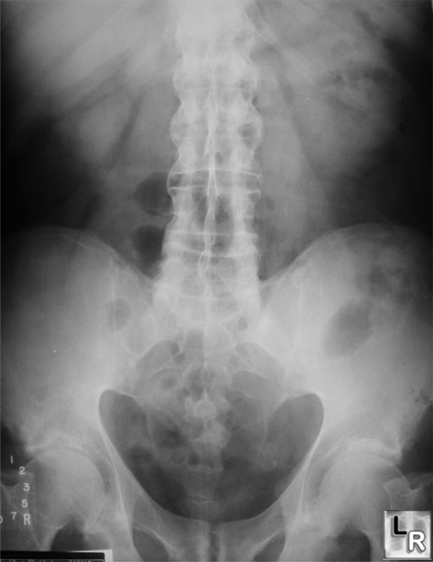
Ankylosing Spondylitis. Note fusion of
both SI joints and thin, symmetrical
syndesmophytes bridging the intervertebral disc spaces.
|
|
|
