|
|
Cerebral Arteriovenous Malformations
Submitted by Anthony Chang, MD
General Considerations
- True AVMs contain at least one enlarged feeding artery and at least one enlarged early draining vein.
- These vessels arise congenitally during fetal life and can be supplied from any cerebral vessel.
- Superficial AVMs may be supplied from the external carotid artery with a dural component.
- The AVM complex begins with the dilated feeding artery to the core/nidus (vascular cluster of entangled vessels) to enlarged draining veins
CT findings
- Vascular tangles are serpiginous and hyperdense without contrast from the blood pool effect.
- AVMs may contain punctate or curvilinear calcification.
- AVMs will enhance
MRI findings
- Curvilinear flow voids
- MRA for mapping
Angiogram
- Should demonstrate the three components of the enlarged feeding artery, core/nidus, and enlarged draining vein.
- Smaller AVMs may simple demonstrate early venous filling during the arterial phase of enhancement
Associated syndromes
- Sturge Weber
- Wyburn Mason
- Klippel-Trenaunay-Weber
- Osler Weber Rendu
Complications
- Hemorrhage
- Steal phenomenon where blood supply is preferentially delivered to AVM at the cost of normal brain parenchyma and can lead to focal neurological symptoms, seizure and focal atrophy.
- Aneurysms can form and become a source of hemorrhage
Treatment
- Endovascular embolization
- Surgery
- Radiation therapy
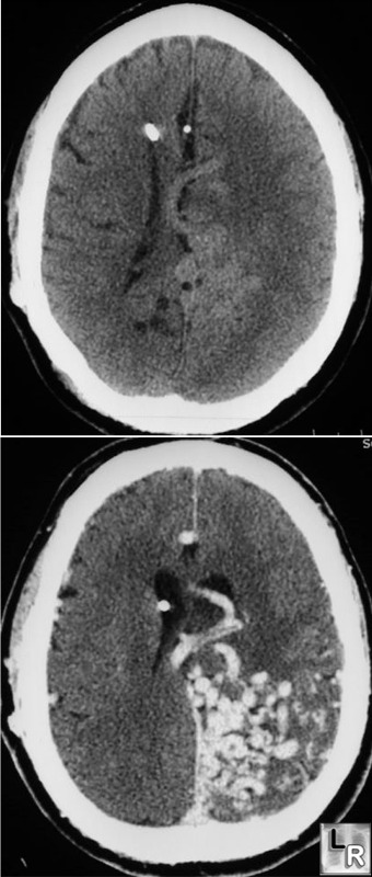
Cerebral Arteriovenous Malformation. Unenhanced (top) and enhanced (bottom) axial CTs of brain shows a
large, serpiginous AVM in the left parietal lobe adjacent to the tentorium.
The Requisites: Neuroradiology 2nd edition, Grossman and Youssem
|
|
|
