|
|
Acute Mesenteric Ischemia
Submitted by Jonathon Dorff, MD
General Considerations
- Acute interruption of blood flow to small or large intestine
Causes:
- Arterial embolism
- Superior mesenteric artery most commonly involved
- Arterial thrombosis
- Nonocclusive mesenteric ischemia
- Low cardiac output state with diffuse mesenteric vasoconstriction
- Mesenteric venous thrombosis
Risk Factors
- Atrial fibrillation/flutter
- Recent acute MI
- Ventricular aneurysm
- Cardiomyopathies
- Valvular disease
- Hypovolemia or hypotension (sepsis)
- Coagulation disorders or malignancy
- Pancreatitis
- Portal hypertension/cirrhosis
- Medications
- Vasopressor medications
- Beta-blockers
- Digoxin
- Diuretics
Clinical signs and symptoms
- Severe abdominal pain out of proportion to physical exam
- Pain initially of a visceral nature and poorly localized
- Nausea
- Vomiting
- Diarrhea
- GI bleeding may be present
Imaging
- Plain abdominal radiographs (abnormal in 20-60% of cases)
- Thumbprinting
- Non-specific finding indicating intestinal wall edema with hemorrhage in the setting of acute mesenteric ischemia
- Pneumatosis
- Portal venous gas
- Pneumoperitoneum
- All are indicative of infarcted bowel
- CT
- Bowel wall thickening from edema or hemorrhage
- Lack of enhancement indicates infarction
- Pneumatosis, portal venous gas, pneumoperitoneum
- Intraluminal thrombus in involved vessel
- Mesenteric angiogram
- Can distinguish between arterial embolic and thrombotic causes of acute mesenteric ischemia
Treatment
- Mesenteric angiogram
- Vasodilator therapy
- Thrombectomy/Embolectomy
- Surgery
- Thrombectomy/Embolectomy
- Arterial bypass
- Resection of necrotic bowel
Complications
- Sepsis/septic shock
- Multiple system organ failure
- Death
Mortality
- 70-90% overall
- From arterial embolism: 60-80%
- From arterial thrombosis: 70-100%
- From nonocclusive mesenteric ischemia: 40%
- From mesenteric venous thrombosis: 25-30%
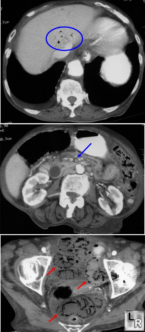
Mesenteric Ischemia. Top CT image shows gas in portal venous system (blue circle);
center image shows absence of contrast
in superior mesenteric artery due to thrombosis of this vessel (blue arrow)
[The patient also has a markedly dilated common duct, not related to
mesenteric ischemia]; lower image shows extensive pneumatosis intestinalis
(red arrows)
|
|
|
