|
|
Cerebellar Hemangioblastoma
- Benign neoplasm
- 80% found in cerebellum
- Remainder in spinal cord > medulla > cerebrum
- Account for 10% of posterior fossa masses (vestibular schwannomas and metastases rule here.)
- Occur in ages 30 to 40
- Relationship to von Hippel-Lindau disease
- 20% occur in patients with von Hippel-Lindau disease (multiple lesions).
- 35-60% of von Hippel-Lindau disease patients have hemangioblastomas
- von Hippel-Lindau disease
- Clinical findings
- Headache
- Ataxia
- Nausea
- Vomiting
- Vertigo
- Polycythemia caused by increased erythropoietin found in 40%.
- Spinal lesions may present with subarachnoid hemorrhage
- Findings on CT and MRI:
- Cystic lesion in the cerebellum with an avidly enhancing mural nodule (75%)
- Purely solid enhancing lesion (10%)
- Enhancing lesion with multiple cystic areas (15%)
- Findings on angiography:
- Vascular nodule within an avascular mass
- Serpentine vessels
- Treated with surgical removal of solid nodule
- Cystic component is not neoplastic
- DDx:
- Similar appearance to Juvenile pilocytic astrocytoma
- But that is typically found in patients 5 to 15 years of age
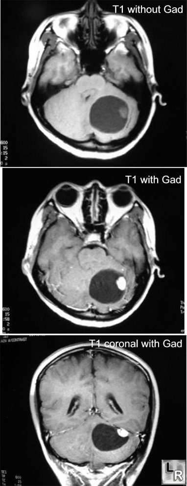
Cerebellar Hemangioblastoma. MRI of brain shows a cystic lesion in the cerebellum
with an enhancing nodule (post-Gadolinium)
|
|
|
